 |
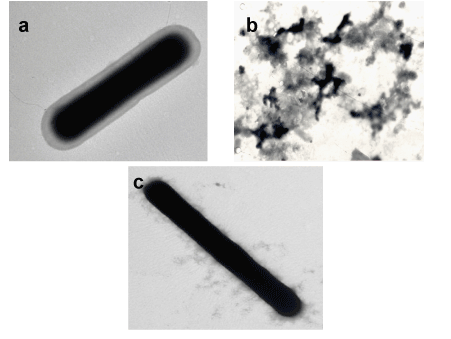
| Figures 1(a-c): Electron micrographs of different sample preparations of C. difficile after (1a) suspension in PBS without matrix; shows an intact cell with a distinct capsule and outer polymeric layers, (1b) exposure to DHB in free solution; shows total lysis, (1c) cells taken off the target plate immediately following the addition of DHB; shows loss of the outer polymeric layers, perhaps due to rapid evaporation of the matrix solvent. |