 |
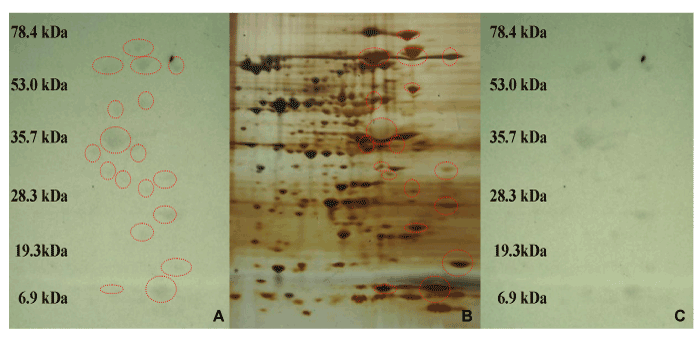
| Figure 3: B. subtilis cell lysates showing protein spots binding radioactive calcium (45CaCl2) and antibody crossreactivity in control cells. Untreated, exponentially growing cells were harvested and lysed as indicated in Materials and Methods. After 2-D gel electrophoresis, proteins were transferred to nitrocellulose and PVDF membranes, to perform 45Ca-autoradiography and Western blots as described in the Materials and Methods section. A) Protein spots, indicated by circles, were identified by 45Ca-autoradiography. B) 2-D gel stained with silver stain showing protein spots identified by both 45Ca2+ autoradiography and EF-hand motif antibody. C) Protein spots identified by 45Ca autoradiography. |