 |
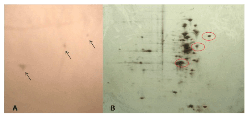
| Figure 4: Western blot analysis of differentially expressed proteins after EGTA treatment. B. subtilis cell lysate proteins were separated by 2-DE and blotted to PVDF as described in Materials and Methods. A) Protein spots crossreacting with the EF-hand antibody. B) 2-D gel stained with silver stain. Red circles indicate the proteins recognized by the antibody. |