 |
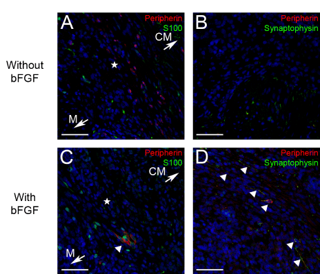
| Figure 3: Transplantation of Enteric Cells with and without bFGF. Approximately 250,000 enteric cells were injected in the rodent stomach with and without bFGF. Peripherin (red) and S100 (green) expression in the injection site without bFGF (A) and with bFGF (C) show the formation of ganglion-like structures in the injection site (star) in the presence of bFGF(triangle, C). Peripherin (red) and synpatophysin (green) expression in the injection site without bFGF (B) and with bFGF (D) show synaptic vesicles are present in these ganglion-like structures (triangle, D) that are formed in the presence of bFGF. The direction of the mucosa (M) and circular muscle (CM) are noted with arrows on A & C and this orientation is consistent throughout the figure. Nuclei are stained with DAPI (blue) and scale bars represent 50 um. |