 |
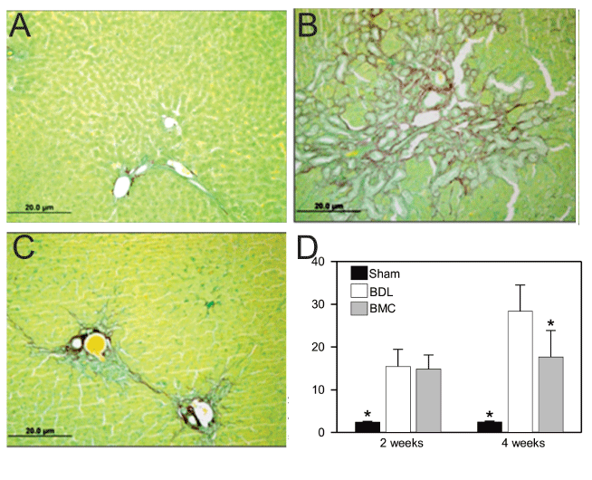
| Figure 3: Extent of collagen deposition in Sham, BDL and BMC groups 4 weeks of BDL. A) Sirius Red staining from liver section from a Sham rat. B) LDB rat C) BMC-treated rat. Magnification 200X. D) Percentage of collagen area quantified as the red-stained area, measured in pixels at 2 and 4 weeks. (*P<0.05 compared with BDL and BMC, **P<0.05 compared with BDL). BDL: Bile duct ligation. BMC: Bone Marrow Cell. |