 |
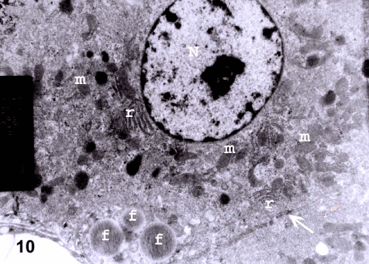
| Figure 10: An electron micrograph from a section in the liver of prophylactic group III showing hepatocyte with euchromatic nucleus (N), regular rough endoplasmic reticulum (r) and mitochondria (m). Cytoplasmic fat globules (f) are noticed. Tight junction (arrow) between two hepatocytes is also noticed (TEM X 11000). |