 |
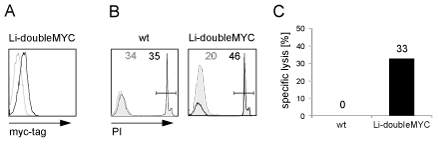
| Figure 4: Myc-tagged human γc expressed on PBLs can be detected by anti-myc ab and mediates complement lysis. (A) PBLs were transduced with human Li-doubleMYC γc, enriched for myc expression by MACS and analyzed by flow cytometry using anti-myc ab. Wt γc-transduced PBLs were used as a control (wt: tinted, Li-doubleMYC: solid black line). (B) CDC assay of wt and myc-tagged human ?c-transduced PBLs was performed by subsequent incubation with anti-myc ab and complement factors (solid line, black numbers), or complement alone as a control (tinted, grey numbers). Dead cells were detected with PI and quantified by flow cytometry. (C) Calculated specific lysis. |