 |
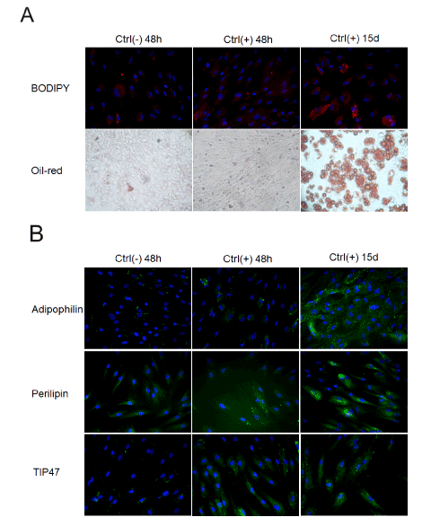
| Figure 1: Visualization of lipid droplets and PAT proteins during the SVF differentiation using BD Pathway Bioimager 855 (20x objective magnification). SVF cells were incubated for 48 hours in adipogenic MDI medium for initiation of differentiation (Ctrl (+) 48 h) and then without differentiation factors for 15 days (Ctrl (+) 15d). The negative control cells (Ctrl (-)) were incubated without differentiation factors. A. Lipid droplets stained using BODIPY 492/503 and Oil-Red-O (10x objective magnification). B The visualization of PAT protein during differentiation (stained using antibody with Cy3). (red- BODIPY, blue-Hoechst, green-PAT proteins). |