 |
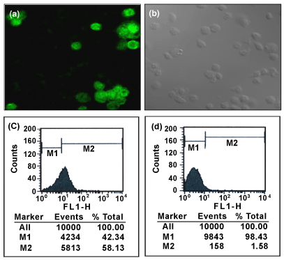
| Figure 2: Immunofluroscence analysis of c-Kit-GFP protein expression in differentiated SSCs at 72 hrs post transfection showing surface expression of c-Kit protein (a). Light microscopic image of SSCs transfected with 1.1 kb control-GFP construct at 72 hrs post transfection (b) (MagnificationX 630). Flowcytometry analysis of c-Kit-GFP protein expression in differentiated SSCs at 72 hrs post transfection with 4.4 kb c-Kit-ORF-GFP insert (c) and 1.1 kb control-GFP insert transfected SSCs at 72 hrs post transfection (d). Figures shown are the representative picture from two different experiments performed on two different occasions. |