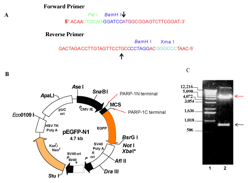
A. Forward primer with PstI and BamHI restriction sites and reverse primer with BamHI and XmaI restriction sites were synthesised by Genset. Arrows in the forward and reverse primers indicate PARP-1 start and PARP-1-250 end site respectively. B. pEGFP vector map taken from Clontech literature is modified to show PARP-1 fragment N-terminal and C-terminal sites. C. PARP- 1-N-terminal fragment was cloned into pEGFPN1 vector and digested with BamH1. Digestion products were analysed by agarose gel electrophoresis with 1kb DNA ladder. Red and black arrows indicate bands corresponding to pEGFPN1 vector at 4700 bp and PARP-1-N-terminal fragment at 750 bp respectively. Data shown is representative of three separate experiments.