 |
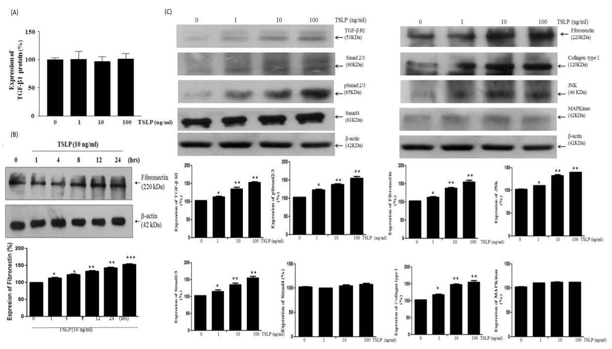
| Figure 2: Effects of TSLP on signal transduction and TGF-β1 protein levels in HFL-1 cells. Cells were cultured in 25 cm2 flasks. Supernatants from conditioned cells were collected and subjected to ELISA for detection of TGF-β1 and to Western blot for detection of fibronectin. Cells were serum starved (0.5% FBS) and treated dose-dependently (0, 1, 10 or 100 ng/ml for 24 hours, Figure 2A) with TSLP, after which ELISA was used for detection of TGF-β1. Alternatively, cells were serum starved (0.5% FBS) and treated time-course-dependently (0, 1, 4, 8, 12, 24 hours, Figure 2B) with TSLP, after which Western blot was used for detection of fibronectin. Cells were lysed and 100 μg of protein lysate was resolved by 7.5% SDS-PAGE followed by Western blot using antibodies against type I receptor TGF-β, pSmad2/3, Smad2/3, Smad4, fibronectin, collagen, JNK, and MAP Kinase. Experiments were repeated three times and consistent results were observed. Experiments were duplicated (n=4). It is evident that TSLP significantly increases type I receptor TGF-β, pSmad2/3, Smad2/3, fibronectin, collagen, and JNK without affecting the secretion of TGF-β1. P*<0.05, P**<0.01 versus control. |