 |
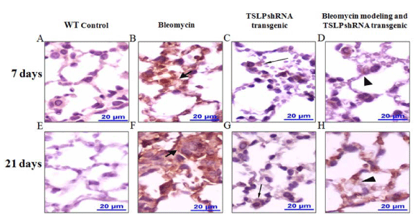
| Figure 6: Immunohistochemistry of bleomycin-treated mice shows intense staining for TSLP proteins in the pulmonary tissues. Regardless of treatment with bleomycin or saline, mice pulmonary tissue samples were perfused, fixed, and embedded in paraffin. Immunohistochemistry was performed according to the protocol listed in the Methods section. A, E: Wild-type mouse pulmonary samples (high power) using IgG control show no brown pigment (i.e., no TSLP expression) at either 7 or 21 days. B, F: Bleomycin-treated mouse pulmonary samples show intense staining for TSLP in the foci of the injured pulmonary tissue; these areas appear to be extracellular (thick arrows). C, G: TSLP shRNA transgenic mouse pulmonary samples show mild TSLP staining associated with type II alveolar cells (thin arrows). D, H: After bleomycin modeling, TSLP shRNA transgenic mouse pulmonary samples have smaller foci of the injured pulmonary tissue. TSLP staining appears to be extracellular (arrow heads). |