 |
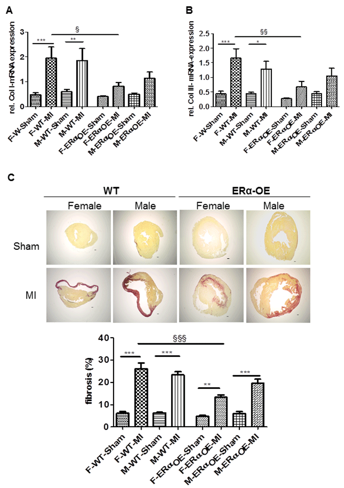
| Figure 6: Cardiomyocyte-specific overexpression of ERα attenuates fibrosis. A-B: Assessment of mRNA expression of fibrosis markers Col I and III in the LV tissues from female and male WT- and ERα-OE mice 2 weeks after sham and MI by qRT-PCR. ERα-OE attenuates the MI-induced expression of Col I and III after MI. Data expressed as mean ±SEM; n ≥ 8 mice per group. § for the genotype effect: females: p<0.05 and p<0.01 for each gene. * for MI effect for each gene: WT-female: p<0.001; ERα-OE-female: p<0.001;WT-male: p<0.01 and ERα-OE-male p<0.05. C: Sirius red staining of representative LV tissue (upper panel, scale bar 200 μm) and fibrosis quantification expressed as the percent of fibrosis area in entire LV cross-section (lower panel) in WT- and ERα-OE mice 2 weeks after sham or MI surgery. The extent of interstitial collagen accumulation in female ERα-OE was significantly less in comparison to female WT-mice after MI. Data expressed as mean ± SEM of 4 to 6 animals per group. § for the genotype effect: p<0.001. * for MI effect (MI vs. sham): WT-female: p<0.001 and ERα-OE-female p<0.01; WT-male: p<0.001 and ERα-OE. |