 |
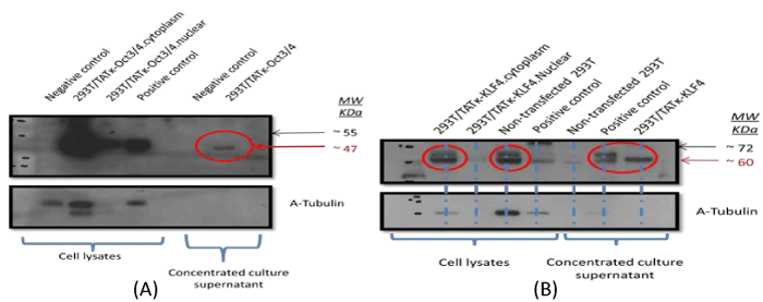
| Figure 5: TATκ fusion protein expression and secretion in stable mixed populations of 293T cells by Western blot analysis. Intracellular expression (cell lysates) and secretion of (a) TATκ-OCT-3/4 (~60 KDa), and (b) TATκ-KLF4 proteins in the concentrated culture medium after 4 weeks of puromycin treatment (2.5 μg/mL). Cell lysates and concentrated culture medium from untransfected 293T were used as negative control. Commercially available mouse embryonal carcinoma (F9) and mouse testis extract cell lysates were used as positive controls. An anti-α-Tubulin antibody was used to detect α-Tubulin (~ 55 KDa), which was used as a loading control for each sample. |