 |
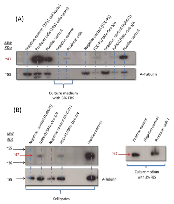
| Figure 9: Oct3/4 protein transduction analysis. A total of 1x10⁵ target cells (JURKAT, FDC-P1 and primary mouse bone marrow cells) were added separately to 2.5 mL of culture medium containing secreted TATκ-Oct3/4 protein (supplemented with 3% heat-inactivated FBS). The cells were further incubated for 3 hours and lysed for Western blot analysis. Bands of the correct size were detected in both JURKAT and FDC-P1 cells, indicating the uptake of TATκ-Oct3/4 protein at (a) 24 hours, and (b) 48 hours after protein transduction. For negative control, target cells were incubated with culture medium of the untransfected 293T cells. A commercially obtained mouse embryonal carcinoma (F9) cell lysate was used as a positive control. The secretion of spTATκ-Oct3/4 protein in cultured medium by 293T producer cells was also confirmed as shown in lane 8. α-Tubulin (~ 55 KDa) was used to detect a tubulin protein which was used as a loading control for each sample. |