 |
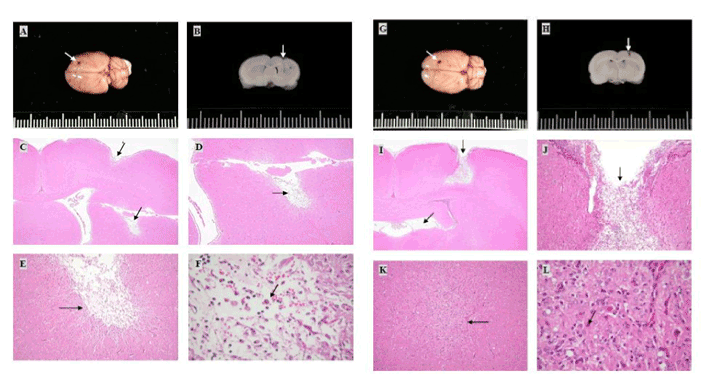
| Figure 2: Gross and histopathological changes in the brain after the rats were grafted with D0 pES (A-F), D12 NP (G-L), and D18 NP (M-R) for 1 month. The sections were stained with hematoxylin and eosin. Black spot lesions were found in the transplantation site of all treatments. White arrows indicate the transplantation sites (A, B, G, H, M, and N). Microscopically, in the D0 pES treatment group, moderate traumatic liquefactive necrosis was noted in the cortex of the brain (C, Magnification×20; D, Magnification×100). Slight granulation with gliosis was found in the lateral ventricle of the brain (E, Magnification×200; F, Magnification ×400) (Black arrows). In the D12 NP group, moderate traumatic liquefactive necrosis with numerous mononuclear cells infiltration was noted in the cortex (I, Magnification×20; J, Magnification×100). Moderate granulation with gliosis was found in the parenchyma of the brain (K, Magnification×100; L, Magnification×400) (Black arrows). In the D18 NP group, moderate traumatic liquefactive necrosis (O, Magnification×20) with menigiogranulation was noted in the cortex of the brain (P, Magnification×400). Slight granulation with gliosis and hemosiderosis were found in the lateral ventricle of the brain (Q, Magnification×100; R, Magnification×400) (Black arrows) |