 |
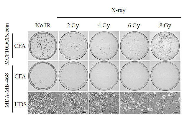
| Figure 2: Representative images of CFA and HDS assay result. CFA or HDS assay was performed for MDA-MB-468 cells, and representative images for each assay were shown in the middle and the bottom panels. For CFA, 900 cells for non-irradiated, 2.4 × 10³ cells for 2 Gy irradiated, 1.2 × 10⁴ cells for 4 Gy irradiated, 3 × 10⁴ cells for 6 Gy irradiated, or 1.5 × 10⁵ cells for 8 Gy irradiated cells were plated onto 60-mm, 100-mm, 150-mm, 150-mm, or 150-mm diameter plastic dishes, respectively. Colonies were fi×ed and stained after 13-day incubation period. For HDS assay, 3 × 10⁵ cells per T25 flask was used for the each dose of ×-ray irradiation. In comparison with MDA-MB-468, the representative images of HDS assay with MCF10DCIS.com were shown in the upper panels. For MCF10DCIS.com, 300 cells for non-irradiated, 800 cells for 2 Gy irradiated, 4800 cells for 4 Gy irradiated, 1.5 × 10⁴ cells for 6 Gy irradiated, or 1.5 × 10⁵ cells for 8 Gy irradiated cells were plated onto 60-mm, 100-mm, 150-mm, 150-mm, or 150-mm diameter plastic dishes, respectively. Scale bar: 100µm |