 |
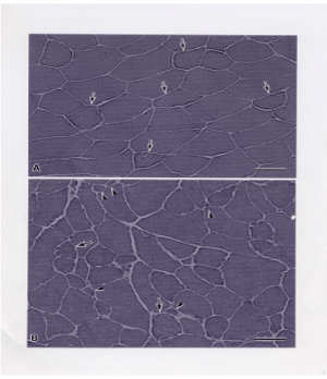
| Figure 1: Hematoxylin and eosin (HE) staining of the cross-sectioned quadriceps femoris muscles of control chow-fed mouse (A) and mouse with diet induced obesity (DIO) (B). In control mouse muscle (A), the scattered myofibers (arrow) with slight granular sarcoplasmic appearance and slit like structures are seen. The DIO muscle (B) contains scattered angulated small diameter myofibers (arrow head) and apparently degenerating myofibers (arrow) in which nonhomogeneous sarcoplasmic appearance, slit like structures, and / or surface indentation are observed. Bar=50 μm (A,B). |