 |
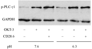
| Figure 2: Phosphorylation of PLC-γ1 stimulated by OKT-3 and CD28.6. After Jurkat cells had been cultured in pH 7.6 and 6.3 media for 24 h, they were stimulated with OKT-3 (0.2 μg/ml) and CD28.6 (5 μg/ml) for 5 min. Whole cell extracts were analyzed using anti-p-PLC-γ1 mAb and anti-GAPDH mAb as described in the legend of Figure 1. |