
|
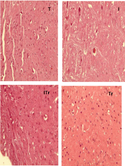
| Figure 3: Microscopic study of cerebral cortex performed by staining (HE) in mice poisoned with AlCl3 orally (100 mg/kg) and intoxicated treated mice (IT) with curcumin for 11 weeks with dose (45 mg/kg) (T) cerebral cortex of control mice (G×400). (I) cerebral cortex characterized by a decrease in cell density and neuronal degeneration (G×400), (IT1) shows a decrease of edema and vacuolation in intoxicated treated (G×400). |