 |
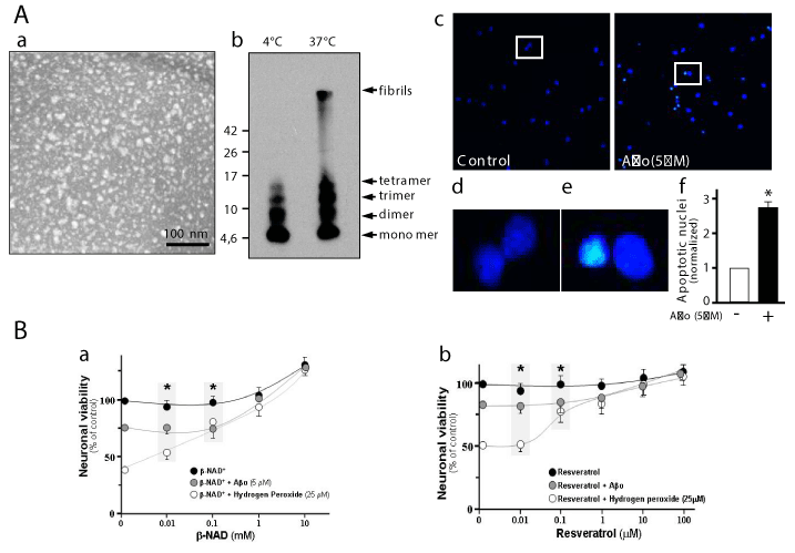
| Figure 1: Effect of SIRT1 modulators on Neuronal viability challenged with Aβo. Aβ peptide was aggregate at 4°C and stained with 2% uranyl acetate and photographed with an electron microscope (A, a), Western blot for 50 μM Aβ aggregated was made, aggregation at 37°C was used as control (b), hippocampal neurons at 14 days in vitro (DIV) control and treated with 5 μM Aβo were stained with Hoechst (c), zoom areas show the normal nuclei and picnotic and condensed nuclei (d), graph show the quantification of apoptotic nuclei in hippocampal neurons (e). (B) Hippocampal neurons at 14 days in vitro (DIV) were treated for 24 h with different concentration of β-NAD+ (0.01-10 mM) (black circle) or β-NAD+ plus 5 μM Aβo (grey circle), 25 μM of Hydrogen Peroxide was used as control positive for neuronal damage (white circle)(a). Resveratrol (Res) (0.01-100 μM) (black circle) or Res plus 5 μM Aβo (grey circle), Hydrogen Peroxide was used as control positive for neuronal damage (white circle) (b), the viability assay was made with MTT reagent. Error bars indicate S.E.M. Fourth experiments were made triplicate, *p *p<0.05 (results enclosed in square). Neurotoxicity for Aβo used in this experiment was defined as a decrease or increase in number of condensed nuclei by quantification of at least 20 different representative areas for each treatment. Scale bar, 100nm. |