 |
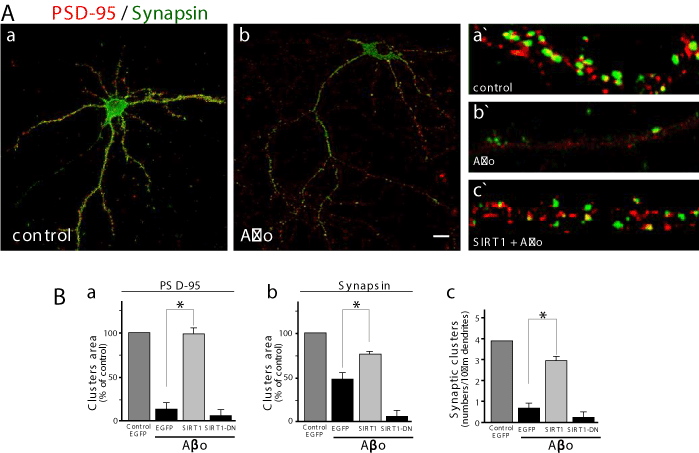
| Figure 3: SIRT1 maintains the integrity of the pre-and post-synaptic markers in hippocampal neurons. Hippocampal neurons at 3 DIV were transfected as shown in Figure 2 (A). Neurons were fixed and PSD-95 and Synapsin was labeled with by immunostaining. Representative images of transfected control neurons with SIRT1/ EGFP, control neurons (a) or challenged wit 5 μM Aβo (b), magnification of the micrographs show pre-(PSD-95, red) or post-sinaptic markers (Synapsin, green) in (a`) control Neurons and then (b`) treated with 5 μM Aβo or (c) SIRT1/EGFP plus 5 μM Aβo. (B) The graphs shows a quantification of the area of clusters and synaptic contacts, (a) area of clusters of PSD-95 (control: first grey bar; Aβo: second black bar; SIRT1 plus Aβo: grey third bar; SIRT1DN plus Aβo: fourth black bar), (b) area of clusters of Synapsin (control: first grey bar; Aβo: second black bar; SIRT1 plus Aβo: third grey bar; and SIRT1DN plus Aβo: fourth black bar), (c) show the synaptic clusters; a pair formed by PSD-95 and Synapsin was quantified (control: first grey bar; Aβo: second black bar; SIRT1 plus Aβo: third grey bar; and SIRT1DN plus Aβo: fourth black bar). Experiments were made triplicate. Error bars indicate S.E.M. n=40-50 transfected neurons and p values were determined by t-student test. *p<0.01. (A) Scale bar: 10 μm |