 |
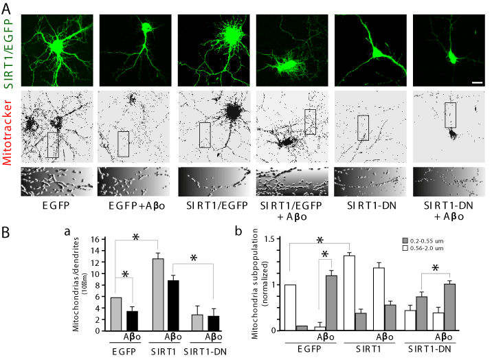
| Figure 4: SIRT1 maintains the integrity of the mitochondria in hippocampal neurons. Hippocampal neurons at 3 DIV were transfected as shown in Figure 2. Mitochondria was stained with orange mitotracker, then fixed and photographed in confocal microscope. (A) Representative images of transfected neurons under different conditions (first row), the neurons transfected were stained with Mitotrackers (second row), a zoom showing the mitochondrial morphology under the treatments is shown (third row). (B) Numbers of mitochondria in transfected neurons was quantified (a), different subpopulation of mitochondria under different conditions was quantified (b). Experiments were made triplicate, p values were determined by Kruskal-Wallis/Dunn (*p<0.01, **p<0.001). Error bars indicate S.E.M. n=40-50 transfected neurons. Scale bar, 10 μm. |