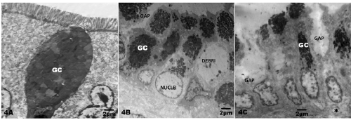
4B. TEM from a 4y old MCMA dog showing a disrupted epithelium with gaps between cells (GAP), wide spaces occupied by cell debris (DEBRI) and enlarged basal cell nuclei (NUCLEI). Goblet cells are marked GC. TEM x 7290
C. TEM from a 5.4 y old MCMA dog showing ample gaps between epithelial cells (GAP) with swollen cytoplasm and vacuolization of basal cells (*).Goblet cells are marked GC TEM x 7290