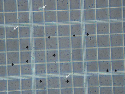
Pluripotent stem cells (white arrows), negatively stained for Trypan blue. Cells have machinery to pump out Trypan blue dye and therefore appear as white “glowing” balls.
Totipotent stem cells (black arrows), positively stained for Trypan blue. Cells lack machinery to pump out Trypan blue dye and therefore appear as dark spherical balls.