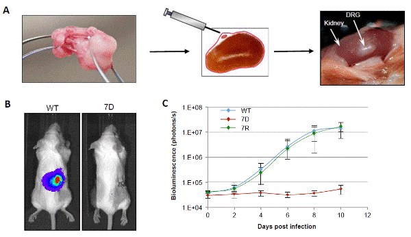
A. Schematic of DRG tissue xenotransplantation. Fetal DRG harvested from spinal cord (left panel) is implanted under the kidney capsule of a SCID mouse (middle panel). After vascularization, the implant is exposed and injected with virus for in vivo growth analysis. B. Image of SCID-hu mouse used for in vivo growth analysis depicting lack of ORF7D VZVLUC growth in a DRG xenotransplant. Image adapted from Zhang et al. (2007). C. In vivo (SCID-hu mice with DRG xenotransplants) growth curve analysis of ORF7D VZVLUC in parallel with WT VZVLUC and ORF7 Rescue (7R) VZVLUC. The DRG was inoculated with 5×10³ PFU WT VZVLUC, ORF7D VZVLUC and ORF7R VZVLUC, as indicated. VZV replication was monitored daily by IVIS for one week as bioluminescence emitting from each DRG was measured. Each line represents an average of the data from 3 different DRG samples, all infected with the same virus.