 |
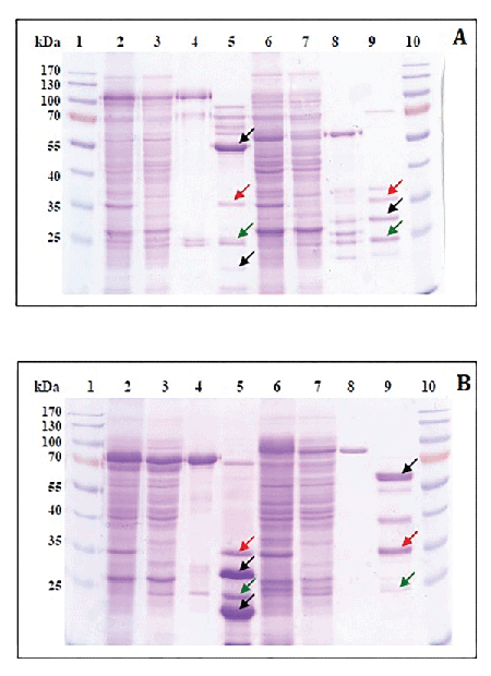
| Figure 1: SDS-PAGE analysis of GBV-C non-structural proteins. A: 1, 10-Ladder (ThermoScientific, USA); 2-bacteria lysate (pGEX-4T2-GBV-C-NS3); 3-supernatant of bacteria lysate (pGEX-4T2-GBV-C-NS3); 4-protein bound to glutathione beads (pGEX-4T2-GBV-C-NS3); 5-glutathione beads cleaved with thrombin (pGEX-4T2-GBV-C-NS3); 6-bacteria lysate (pGEX-4T2- GBV-C-NS4A/B); 7-supernatant of bacteria lysate (pGEX-4T2- GBV-C-NS4A/B); 8-protein bound to glutathione beads (pGEX-4T2-GBV-C-NS4A/B); 9-glutathione beads cleaved with thrombin (pGEX-4T2-GBV-C-NS4A/B). B: 1, 10-Ladder; 2- bacteria lysate (pGEX-4T2-GBV-C-NS5A); 3-supernatant of bacteria lysate (pGEX-4T2-GBV-C-NS5A); 4-protein bound to glutathione beads (pGEX-4T2-GBV-C-NS5A); 5-glutathione beads cleaved with thrombin (pGEX-4T2-GBV-C-NS5A); 6-bacteria lysate (pGEX-4T2-GBV-C-NS5B); 7-supernatant of bacteria lysate (pGEX-4T2-GBV-C-NS5B); 8-protein bound to glutathione beads (pGEX-4T2-GBV-C-NS5B); 9-glutathione beads cleaved with thrombin (pGEX-4T2-GBV-C-NS5B). Black arrows indicate recombinant proteins; red arrows indicate thrombin; green arrows indicate GST protein. |