 |
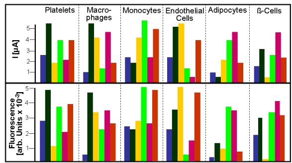
| Figure 4: Determination of signatures specific for EMVs released into the blood from different types of blood and tissue cells by NP-based biosensing (upper panel) and NTA (lower panel) with measurement of current (upper panel) and fluorescence (lower panel) as the read-outs and using the same six types of NPs indicated by the distinct colors. The signatures obtained with the two read-outs are very similar with regard to the relative strength of the interactions between the individual NP types and EMV subspecies, but differ in quantitative fashion. Thus biosensing with both read-outs will reliably provide signatures that unequivocally identify the subspecies and cellular origin of EMVs in the blood (see text for details). |