 |
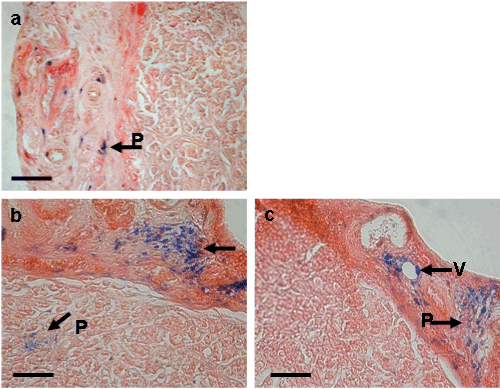
| Figure 6: Histological analysis of tumor slices by optical microscopy after iron oxide staining. The tumors were taken from mice injected weekly by RUMFLs (0.15 μmoles RU (4.3mg/week/kg) either without (a) or in the presence of a 0.44-T magnet (155 T/m magnetic field gradient) placed at the tumor location (b,c). The sections were stained with eosine/saffron for cell visualization and 2% w/v potassium ferrocyanide in acid medium (perls blue solution) for iron detection. Note the presence of iron oxide characteristic of liposome content viewed inside the blood vessels (V) and in the parenchyma (P). Black bars represent 50 mm. |