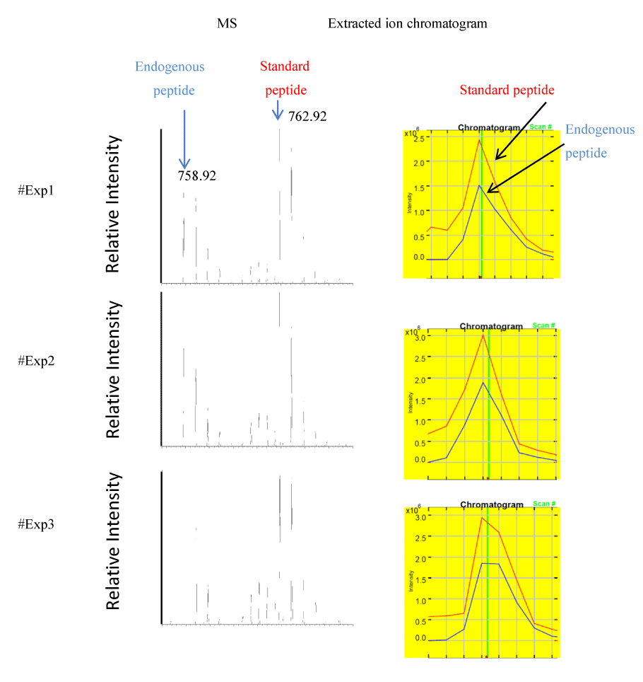
Three aliquots of 50 μg total muscle protein extract were separated by SDS-PAGE as described in Methods. The migration area corresponding to dystrophin was excised, in-gel digested and spiked with optimal concentration (30 nM) of stable isotope labeled standard peptides. The left panel shows MS spectra of unlabeled peptide [IFLTEQPLEGLEK] generated from endogenous dystrophin and the spike-in stable isotope labeled standard peptide [IFLTEQPLEGLEK*]. Both peptides were detected as doubly charged ions at their corresponding m/z value of 758.92 and 762.92, respectively. The right panel shows extracted ion chromatograms used for ratio measurement of unlabeled endogenous dystrophin peptide (blue trace) to the stable isotope labeled standard peptide (red trace).