


 3 / 14

Page 35
conferenceseries
.com
Volume 11
Journal of Proteomics & Bioinformatics Open Access
Computational Biology 2018
September 05-06, 2018
September 05-06, 2018 Tokyo, Japan
International Conference on
Computational Biology and Bioinformatics
Hao Zhu, J Proteomics Bioinform 2018, Volume 11
DOI: 10.4172/0974-276X-C1-113
Reconstructed signaling events unveil how growth and growth arrest are controlled by PCPand Hippo
signaling
Hao Zhu
Southern Medical University, China
H
ow growth is controlled in normal tissues and organs but
not in cancers is a question that has drawn wide attention
but remained poorly understood. To uncover feedbacks and
their properties in control mechanisms consisting of genes and
their products, quantitative methods are needed but not enough,
because gene expression and protein interaction are discrete
events. A new model combining features of lattice models and
vertex models and integrating differential equations, molecular
signaling and mechanical force is developed to investigate growth
and growth arrest of
Drosophila
wing. The model includes key
elements in the Wnt, PCP and Hippo pathways, encapsulates
proteins and their attributes and behaviors into objects, uses
message-passing between objects to simulate signaling between
proteins, simulates cell divisions in a 2D lattice, computes protein
concentrations and cell polarity, captures the spatiotemporal
distribution of signaling events and computes the spatiotemporal
distribution of mechanical stress. The distributions of protein
concentrations, cell polarity, cell division rates, and cell population
agree well with experimental observations, justifying the unveiled
control mechanism. Reconstructed signaling events uncover two
intercellular feedbacks that jointly control growth, patterning
and growth arrest. Specifically, the results indicate that the Warts
repressing Yorkie event (
Wts_Rep_Yki
) distributes densely at the
central in early stages but densely at the periphery in later stages.
The spatiotemporal distribution of this critical event provides the
unprecedented and most pertinent evidence suggesting that growth
is gradually refrained from the periphery to the central of the wing pouch, which is a new and somewhat counter intuitive
finding. The methods can be applied widely to systematically investigating signaling and patterning across the gene, molecular,
cell and tissue levels.
Biography
Hao Zhu has obtained his MS in Computer Science from National University of Defense Technology, Changsha, China and PhD in Pathophysiology from Southern
Medical University, Guangzhou, China. He has completed his Postdoctoral studies in Bioinformatics Institute of Singapore and School of Mathematical Sciences,
University of Nottingham, UK. With a profound interest in Evo-Devo, currently he is working on mechanisms that make new genes (especially lncRNA genes)
function coordinately with old ones to regulate gene expression and control developmental signaling and patterning. He developed a cellular automata style
modeling tool to effectively address signaling events and lncRNA analysis tool/platform (Long Target) to analyze the evolution and function of lncRNAs.
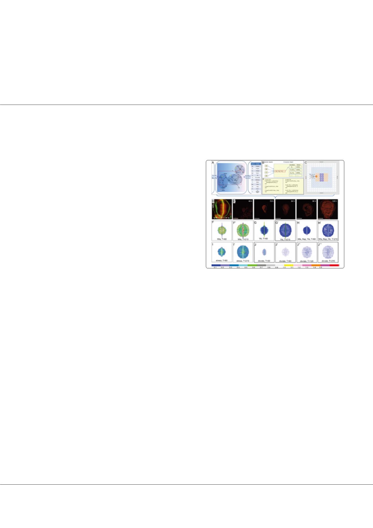
zhuhao@smu.edu.cnStructure and results of the wing pouch growth model. (A) The model
contains key elements in the Wnt, PCP and Hippo pathways. (B) Proteins
are encapsulated into objects and signaling between proteins is captured
as message-passing between objects. (C) In a 2D lattice each unit contains
a set of PDE/ODE and represents a wing cell. Initial cells (the purple and
pink ones) are at the central, new cells (the blue ones) generated by cell
division make the cell population grow. (D) The wild-type of Wg and Dll
distributions (note the two outer green ribbons are not wing pouch). (E)
The wild-type of growth process. (FG) Wts and Yki concentrations in the
lattice at early and late stages (T indicates non-dimensionalized time).
(HI) The Wts_Rep_Yki event and cell stress in the lattice at early and
late stages. (J) Cell division rates in cells decrease (the blue color darkens)
gradually, making growth finally arrested. The color bar indicates non-
dimensional protein concentrations.