


 4 / 19

Page 33
conferenceseries
.com
June 19-20, 2017 Philadelphia, USA
14
th
International Conference on
Clinical and Experimental Dermatology
Volume 8, Issue 4 (Suppl)
J Clin Exp Dermatol Res, an open access journal
ISSN:2155-9554
Dermatology 2017
June 19-20, 2017
Terahertz multispectral reconstructive imaging of biological specimen
Anis Rahman
1
, Babar Rao
2
and
Aunik Rahman
1
1
Rutgers University, USA
2
Applied Research & Photonics, USA
T
erahertz multispectral reconstructive imaging is an effective tool for soft tissue imaging without any radiation damage
kike X-ray. Here, examples of biological tissue imaging are outlined to elucidate the technique. Reconstructive imaging
utilizes the technique of rasterizing a specimen over a given area. ARP’s instrument allows the T-ray beam to be focused on
a given layer under the surface; therefore, a 3D volume may be rasterized on a layer by layer basis. The reflected intensity is
recorded preserving the exact coordinates over which measurements are done. The intensity matrix is then converted to image
via inverse gridding algorithm. The algorithm is capable of accurate representation of the measured object similar to a charged
couple device as has been explained previously. Here we present images of human skin under different diseased conditions as
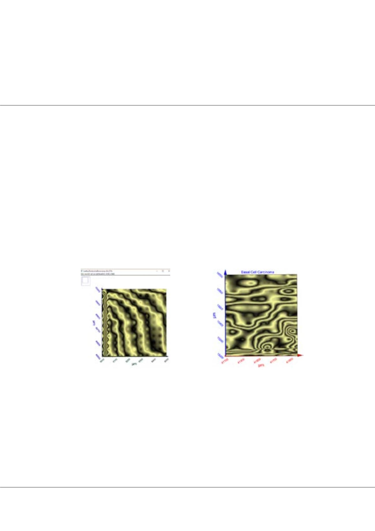
compared with healthy skin samples. Fig. 1(a) exhibits terahertz reconstructive images of a healthy skin sample where regular
cellular pattern is visible. This is expected from the healthy skin tissue. Fig. 1(b) shows an image of a skin sample diagnosed
for basal cell carcinoma. As evident from Fig. 1(b), diseased skin sample has lost its regular cellular pattern which is present
for the healthy skin sample. This lack of systematic cellular structure may serve as an easy visual means to indicate that there
is something wrong with the sample. As outlined in reference [1], a combination of presence or absence of regular cellular
structure, terahertz spectral comparison, and lack or presence or layering information is expected to serve as a fool proof
diagnostic tool for different kind of skin cancers.
Figure 1: (a): terahertz image of healthy skin tissue. (b): Image of skin tissue diagnosed for basal cell carcinoma showing distorted cell structure
Biography
Anis Rahman is an acclaimed Scientist in the field of Nanotechnology. He is a winner of many scientific awards including NASA Nanotech Brief’s “Nano-50” award
twice; CLEO/Laser Focus World’s “Innovation award”. He is the Founder of a terahertz company in Harrisburg, Pennsylvania. He is a recognized Scientific Leader
and Member of professional organizations including the American Chemical Society (ACS), The Optical Society of America (senior member), and the SPIE. He is
the current Chair of Small Chemical Businesses Division of the ACS. He has been an author and co-author of more than 120 papers in peer reviewed journals and
conference proceedings.
a.rahman@arphotonics.netAnis Rahman et al., J Clin Exp Dermatol Res 2017, 8:4 (Suppl)
DOI: 10.4172/2155-9554-C1-059