


 8 / 36

Page 34
Notes:
conferenceseries
.com
Joint Conference
July 17-18, 2017 Chicago, USA
International Conference on
DIAMOND AND CARBON MATERIALS & GRAPHENE AND SEMICONDUCTORS
Volume 6, Issue 6 (Suppl)
J Material Sci Eng, an open access journal
ISSN: 2169-0022
Diamond and Carbon 2017 & Graphene 2017
July 17-18, 2017
Suspended graphene and nanoscrolls explored by nanofocused x-rays
Gilbert Chahine
and
Johann Coraux
The National Centre for Scientific Research, France
S
tructure determination of crystal lattice parameters and orientation with high precision is rather straightforward for bulk
3D-materials. X-ray diffraction proved especially powerful in this respect across the years. On the contrary, structural
determination of 2D crystals with the help of x-rays is more demanding. So far, exploiting interferences between a crystalline
substrate and graphene, it was possible to accurately determine the lattice parameter of graphene, averaged across the
~1cm2 surface of a sample. Such studies are however restricted to graphene samples of macroscopically uniform crystalline
orientation. However most graphene samples of relevance for potential applications (micro-electronics, telecom, displays)
exhibit in homogeneities, as they are composed of single-crystal grains (~10µm), each having different crystalline orientation
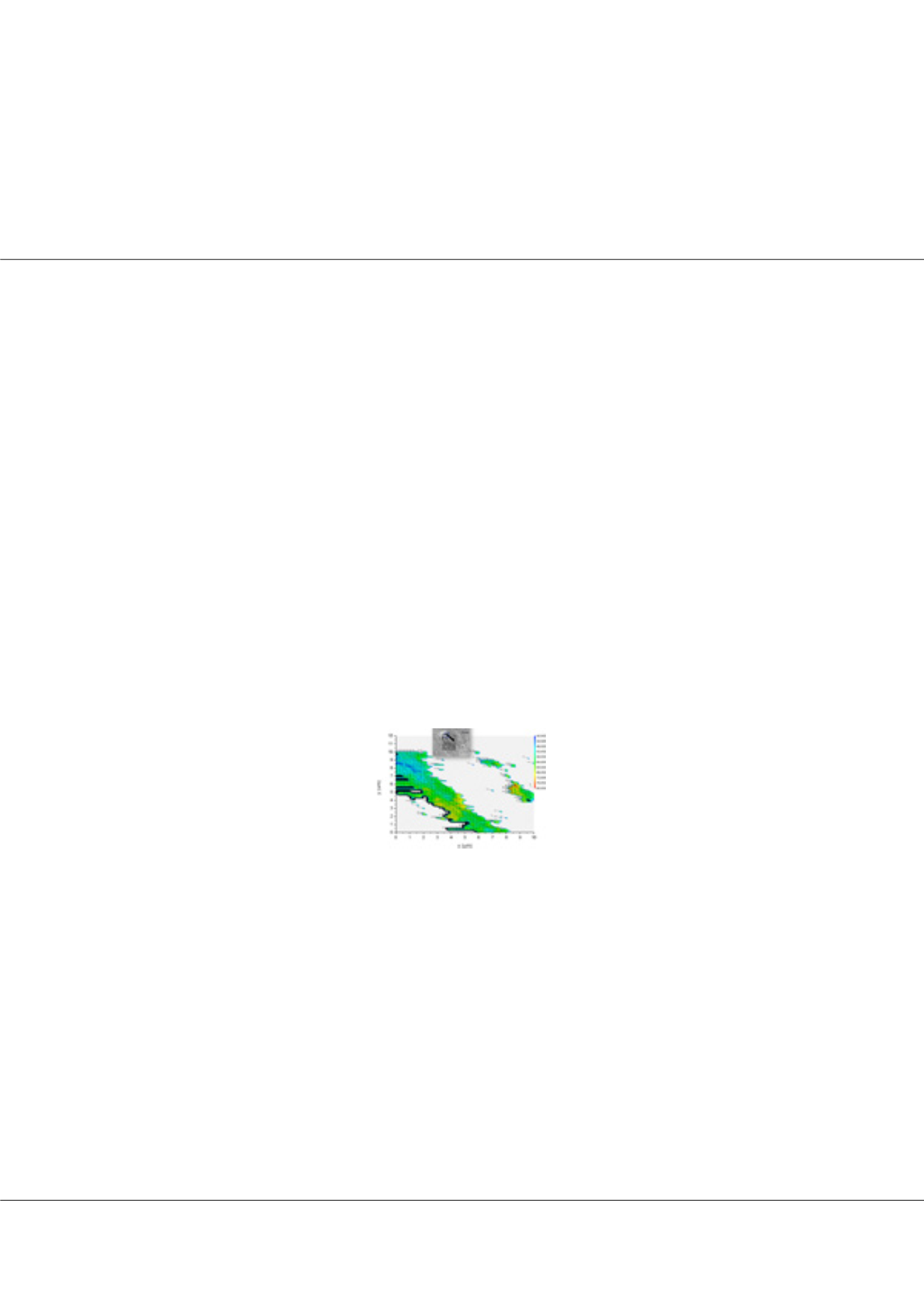
and strain. Finely characterizing such structural features requires probing suspended graphene with the help of nano-shaped
beams. Using, electron beams instead of nanofocused x-rays, we managed to conduct simultaneously small and wide angle
x-ray scattering (SAXS/WAXS) characterizations with high resolution in reciprocal space and an unprecedented resolution
of 200nm in real space, Accordingly we were able to map the structural variations in two- dimensions, revealing in this way
strain maps for the suspended few-layer graphene membrane and the morphological features at the edges of the flakes, where it
forms scrolls with a typical length of the order of 10µm and a diameter of the order of 10nm. The orientation of the nanoscrolls
could for instance be resolved. Our complementary analysis with spatially-resolved Raman spectroscopy provides the unique
opportunity to unambiguously determine the Grüneisen parameters of graphene, linking the deformation to the energy of its
vibration modes, without any particular assumption. These experiments pave the way to advance
in-situ
experiments and for
exploring 2D crystals and their phase transitions using synchrotron radiation especially with the future upgrade programs in
the European Synchrotron for outstanding expected brilliance.
Biography
Gilbert Chahine is a Research-Engineer at the CNRS and working on the BM02 beamline at the European Synchrotron (ESRF) in Grenoble France. After a PhD
in Materials Science with the highest distinction degree at the CEA in France, during his Post-doc he developed a new x-ray imaging technique (KMap) along with
user-friendly software for the analysis of 5D data sets. This technique is now highly requested by a large community of researchers form international institutes to
perform new types of experiments such as in situ and operando strain imaging, with the highest available resolution, of nano devices for photonics, photo voltaics
and optoelectronics. Besides coordinating several projects involving international academic and industrial institutions in the field of strained semiconductors, he
became interested in adapting the latest advances in synchrotron x-ray sources for a direct model-free in-depth probe of 2D materials’ local structure for a better
understanding of their properties.
gilbert.chahine@esrf.frGilbert Chahine et al., J Material Sci Eng 2017, 6:6(Suppl)
DOI: 10.4172/2169-0022-C1-076