


 16 / 22

Volume 6
Journal of Oral Hygiene & Health
Oral Health Meet 2018
November 29-30, 2018
Page 37
Notes:
conference
series
.com
November 29-30, 2018 Bali, Indonesia
International Conference on
Oral Health and Dental Medicine
Priyanka Tiwari, J Oral Hyg Health 2018, Volume 6
DOI: 10.4172/2332-0702-C2-011
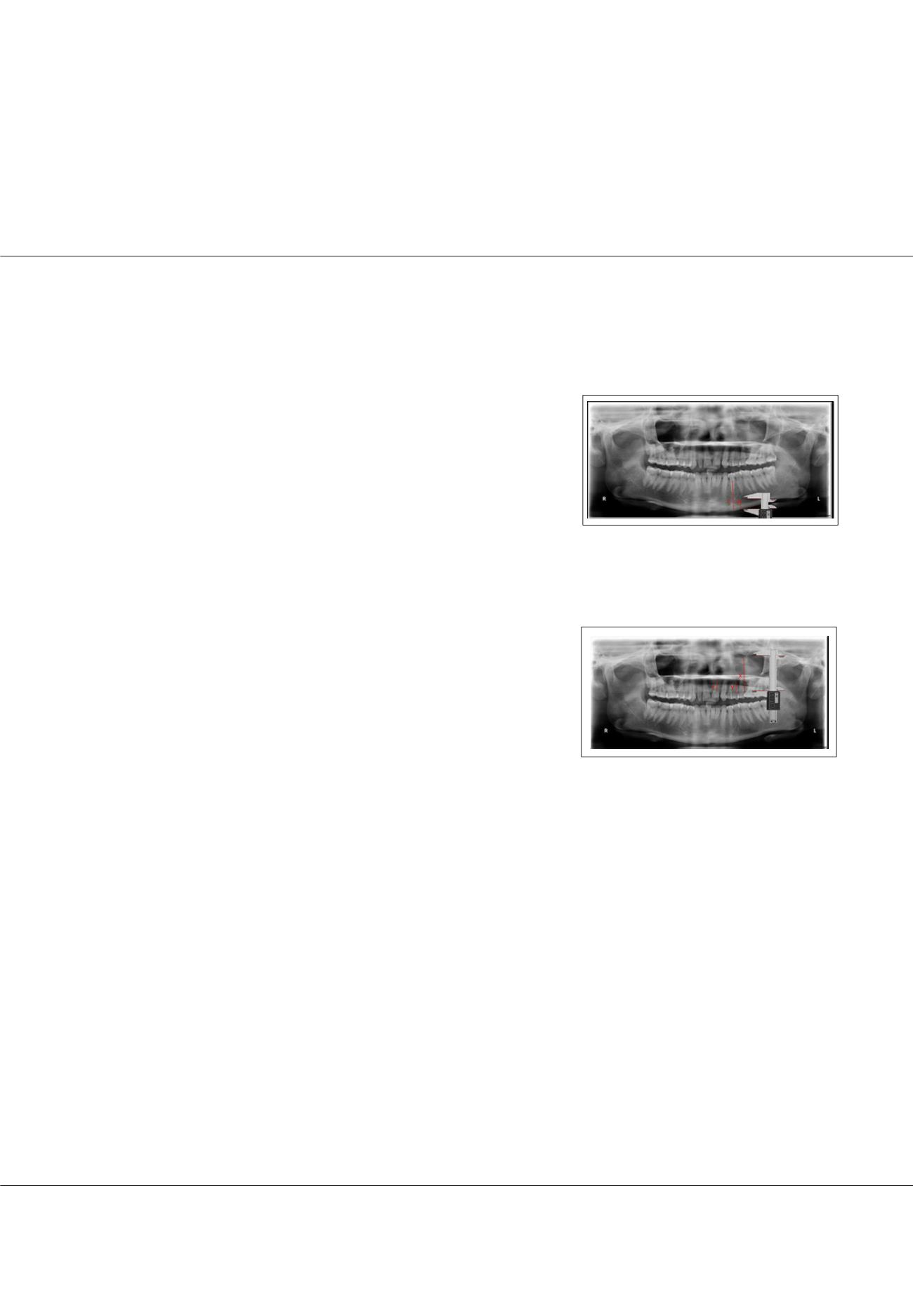
Figure 1:
Mandibular landmarks -‘c’: is
from the inferior border of the mandible
to the alveolar crest. ‘a’: is from the inferior
border of the mandible to lower edge of the
mental foramen in dentulous mandible.
Use of panoramic radiographs for evaluation of maxillary and mandibular residual ridge resorption:
In vitro study
Priyanka Tiwari
Dental Surgeon (Prosthodontist), Malaysia
Introduction:
Ridge resorption is the major reason of mandibular complete
denture losing its stability and retention. The location of the mental foramen can
be identified easily on panoramic radiographs, and radiographic examinations are
considered an important component of Prosthodontics diagnostic and treatment
planning. Also, the location of maxillary landmark is important to known how
much resorption is there. Aim: To determine the average ratio of bone height with
nearest constant anatomical landmarks in maxilla and in mandible. OBJECTIVE-
To find out the association between radiographic findings & prosthodontics, such
as measurements of the amount of resorption and the variation in the treatment
planning of edentulous patients.
Methods:
In this study OPG machine- Kodak C 8000 and Software to calculate the
distance- Screen calipers v2.1 is used. 100 patients OPG was taken in this study.
Major inclusion parameters included were presence of mandibular premolars and
molars, minimal ridge resorption and clear radiographic landmarks visible on the
OPG. After which the landmarks and specific structures were marked on the OPG.
Then measurement was done for distance ‘c’, ‘a’, ‘x’, ‘y’, ‘z’. Lastly, the calculations
from measurements were done to calculate the ratio of c/a, to calculate the ratio of
x/y, to calculate the ratio of x/z.
Result:
The descriptive statistics was done. The C/a ratio mean is 2.71 ± 0.31. The
X/Y ratio mean is 1.49 ± 0.34 and the X/Z ratio mean is 1.51 ± 0.24.
Conclusion:
This ratio can be assessed in edentulous patients and then their further
treatment plan can be decided according to the ratio. The implant placement can be
assessed by using the measurements in this study. KEYWORDS- Average alveolar
bone, panoramic radiograph, mental foramen, mandibular ridge, zygomatic
process, maxillary ridge.
Biography
Priyanka Tiwari, a young & dynamic Indian doctor who has done her BDS- bachelor's in dental surgery
from People's dental academy, Bhopal, (M.P) India and done her MDS- master's in dental surgery in
Prosthodontics from K.M. Shah Dental college & Hospital, Vadodara, (Gujarat) India. She has 5 and half years of clinical experience working in eminent hospitals,
clinics, and college. She practices prosthodontics and general dentistry as well. She has 5 publications in international journals and has done 3 researches. She is
a part of renowned dental associations. Currently settled in Malaysia.
drpriyankatiwari1@gmail.comFigure 2:
Maxillary landmarks- ‘x’:
from line joining most inferior points
of borders of bony orbits to line joining
inferior margins of images of zygomatic
processes. ‘y’: point from zygomatic
process to alveolar crest in maxillary first
molar regions) in dentulous maxilla. ‘z’:
point from zygomatic process to alveolar
crest in maxillary lateral incisor region) in
dentulous maxilla.