| Research Article |
Open Access |
|
| Hanifi BayaroÄßulları1, Vefik Arıca2*, Ali Türkay3, Seçil Arıca4, Yeliz BeyoÄßlu1 and Ramazan Davran1 |
| 1Mustafa Kemal University Medical Faculty, Radiology of Department, Hatay, Turkey |
| 2Mustafa Kemal University Medical Faculty, Pediatric of Department, Hatay, Turkey |
| 3Hatay Children Hospital, Pediatric Clinic, Hatay, Turkey |
| 4Mustafa Kemal University Medical Faculty, Family Medicine of Department, Hatay, Turkey |
| *Corresponding author: |
Vefik Arica, M.D
Medical Faculty of the Mustafa Kemal University
31100, Serinyol, Antakya
Hatay, Turkey
Tel: +90 326 2291000
Fax: +90 326 2455654
Mobile: +90 505 6797877
E-mail: vefikarica@hotmail.com |
|
| |
| Received March 12, 2012; Published June 26, 2012 |
| |
| Citation: BayaroÄßulları H, Arıca V, Türkay A, Arıca S, BeyoÄßlu Y, et al. (2012) Fetal MRI Urography and Antenatal Diagnosis of Posterior Urethral Valve. 1: 113.doi:10.4172/scientificreports.113 |
| |
| Copyright: © 2012 Bayarogullari H, et al. This is an open-access article distributed under the terms of the Creative Commons Attribution License, which permits unrestricted use, distribution, and reproduction in any medium, provided the original author and source are credited. |
| |
| Abstract |
| |
| Posterior Urethral Valve (PUV) is the most common cause of lower urinary tract obstruction in male fetus at the antenatal period. Keyhole sign of urethra, distention of bladder caused by the obstruction and bilateral ureters and pelvicalyceal dilatation can be determined secondary to vesicoureteral reflux. The degree and duration of obstruction determine the prognosis and kidney damage. At antenatal period ultrasound (USG) is the first and the most widely used imaging modality in diagnosis of PUV. As a complementary and an ancillary method MRI is preferred in both maternal and fetal causes. Our case shows the contribution of MRI in the diagnosis of PUV. |
| |
| Keywords |
| |
| Posterior urethral valve; Fetal MRI urography; Antenatal hydronephrosis |
| |
| Case Report |
| |
| The pregnant who was 23 years old with her first pregnancy, according to her last menstrual period she was 19 weeks pregnant and admitted to our hospital with the diagnosis of cystic mass in the fetal abdomen according to the ultrasound which was performed in another institution. Clinical and laboratory examinations of the pregnant had no prominent feature. According to the USG examination which was performed in our clinic using a Siemens Antares 0.5 device, the age of fetus was same with the calculated gestational age according to last menstrual period of mother and the male fetus was at 19 weeks of gestation. The amount of amniotic fluid revealed normal. Pelvicalyceal ectasia of kidneys and distension of bladder were observed (Figure 1). Preliminary diagnosis was considered as a PUV. Because of the bladder distention and the fetal position we had difficulties in detailed visualization of the fetal abdomen and a fetal MR urography examination was planned. We used Philips Achieva 1.5 tesla, XL-torso coil and in T2-weighted single-shot TSE SPIR sequence of the investigation bilateral pelvicalyceal ectasia and bladder distention with bilateral dilatation and tortuosity of ureters and dilatation and elongation of the proximal urethra (keyhole sign) were observed (Figure 2 and Figure 3). Infravesical obstruction was diagnosed secondary to PUV. |
| |
|
|
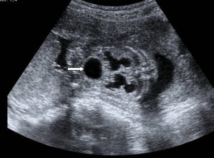
Figure1: USG examination. The pelvicalicsiel dilatation of the kidneys and over-distention of bladder (arrow). |
|
| |
|
|
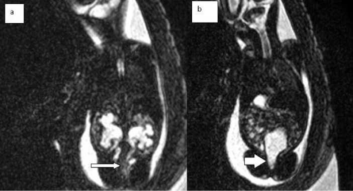
Figure 2: a-b. T2 weighted coronal MR imaging. a- the pelvicalyceal dilatation of the kidneys(arrow), bilateral tortuze and dilatation of ureters(arrow). b- keyhole sing of the proximal urethra (arrow). |
|
| |
|
|
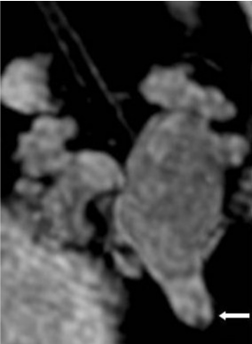
Figure 3: 3D MRÄ° Urography. Bilateral pelvicalyceal ectasia of kidney, bladder distention, key hole sing of proximal urethra (arrow). |
|
| |
| Introduction |
| |
| Posterior urethral valve is the most common cause of lower urinary tract obstruction A PUV is a congenital obstruction caused by a malformation of the posterior urethra. The significance of this obstruction depends on the secondary effects on the bladder, ureters, and kidneys. It may cause irreversible pathologies both on kidney and bladder function. |
| |
| PUV has an estimated incidence of one in 5000 to 8000 in the community, among the only male fetuses [1]. PUV is divided into three types by Young as type 1, type 2 and type 3. Type 1 is the most common seen, as 90 % [2]. |
| |
| As embryologically, PUV is thought to occur as a result of the abnormal insertion of mesonephric duct into fetal cloaca. Valve consists at the level of verumontanum in posterior urethra that prevents urine flow. Clinical signs are the same at tree types of PUV, but severity of symptoms varies depending on the degree of obstruction. |
| |
| Secondary to mechanical obstruction, dilatation and elongation of the proximal urethra, distension of the bladder and VUR develop at the PUV [3]. PUV patients can present ureterohydronephrosis as a consequence of functional (“valve bladder”) or mechanical obstruction and only ~50% of the patients VUR. VUR-mediated dilation observed in the ureters. Hydronephrotic and dysplastic changes of kidneys, subcapsular and perirenal urinoma with the finding of ascites is seen [4,5]. Oligohydramnios and lung hypoplasia are the poor prognosis criteria. Kidney and bladder damage occur depending on the severity and intrauterine detection time of the obstruction. The detection of PUV before 24 weeks extent pressure of obstruction to the process of delivery and worse prognosis [6], the main aim of treatment is to minimize the load on the bladder and kidney, but one-third of these patients, although the most advanced treatments fall in kidney failure to the long-term [7]. |
| |
| Ultrasound is the most widely used imaging modality the diagnosis of PUV and intrauterine fetal examination, because of noninvasive and real time imaging. Maternal obesity, fetal position and oligohydroamnios cause difficulties to examine with ultrasound [8-11]. In such cases, the search for additional fetal anomalies, MRI can be used as the complementary of ultrasound and to obtain additional information. Ultrafast pulse sequences, magnetic resonance (MR) can now acquire high-quality fetal images in an extraordinarily short time, thereby eliminating artifacts due to fetal movements. The lack of ionizing radiation is an advantage of MR, and no side effects for the embryo have been reported [12,13]. There are some studies with MRI in the diagnosis of PUV [14-16]. |
| |
| In our case, in USG examination, remarkable dilatation of the pelvicalyceal structures and over-distention of bladder have been shown. Ureters, due to extensive distension of bladder and proximal urethra and because of the position of the fetus could not be evaluated with USG. |
| |
| In our patient, amniotic fluid was within normal limits and neither increased echogenicity nor cystic lesion in kidneys is evaluated. According to the USG examination the initial diagnosis of our patient was PUV; to obtain certain diagnosis MRI examinations were performed because of the evaluation difficulties with USG. Remarkable pelvicalyceal dilatation of kidneys and over-distention of bladder were observed with MRI, in addition to these findings, tortuozite and dilated bilateral ureters and proximal urethra dilatation (key hole sing) were evaluated. Due to all these findings certain diagnosis of our patient was PUV. |
| |
| Antenatal fetal ultrasound is the first and most important imaging method to examine urinary tract pathologies. The matter of arising from fetus and mother, MRI can be used as a complementary and useful method especially in uncertain diagnosis. Early diagnosis and early intervention can be made. |
| |
| |
| References |
| |
- Cendron M, D'Alton ME, Crombleholme TM (1994) Prenatal diagnosis and management of the fetus with hydronephrosis. Semin Perinatol 18: 163-181.
- Young HH, Frontz WA, Baldwin JC (2002) Congenital obstruction of the posterior urethra. J Urol, 3: 289-365, 1919. J Urol 167: 265-267.
- Cuckow PM, Dinneen MD, Risdon RA, Ransley PG, Duffy PG (1997) Long-term renal function in the posterior urethral valves, unilateral reflux and renal dysplasia syndrome. J Urol 158: 1004-1007.
- Chatterjee SK, Banerjee S, Basak D, Basu AK, Chakravarti AK, et al. (2001) Posterior urethral valves: the scenario in a developing center. Pediatr Surg Int 17: 2-7.
- Macpherson RI, Leithiser RE, Gordon L, Turner WR (1986) Posterior urethral valves: an update and review. Radiographics 6: 753-791.
- Panicker R, Grewal DS (2010) Antenatally diagnosed posterior urethral valves: A dilemma. Medical Journal Armed Forces India 66: 167-169.
- Naghizadeh S, Kefi A, Dogan HS, Burgu B, Akdogan B, et al. (2005) Effectiveness of oral desmopressin therapy in posterior urethral valve patients with polyuria and detection of factors affecting the therapy. Eur Urol 48: 819-825.
- Garel C, Brisse H, Sebag G, Elmaleh M, Oury JF, et al. (1998) Magnetic resonance imaging of the fetus. Pediatr Radiol 28: 201-211.
- Levine D, Goldstein RB, Callen PW, Damato N, Kilpatrick S (1997) The effect of oligohydramnios on detection of fetal anomalies with sonography. AJR Am J Roentgenol 168: 1609-1611.
- Twining P (2003) Genitourinary malformations. In: Nyberg DA, Mc-Gahan JP, Pretrorius DH, Pilu G, Eds. Diagnostic imaging of fetal anomalies. Philadelphia, Lippincott Williams Wilkins p603–660.
- Dinneen MD, Dhillon HK, Ward HC, Duffy PG, Ransley PG (1993) Antenatal diagnosis of posterior urethral valves. Br J Urol 72: 364-369.
- Beers GJ (1989) Biological effects of weak electromagnetic fields from 0 Hz to 200 MHz: a survey of the literature with special emphasis on possible magnetic resonance effects. Magn Reson Imaging 7: 309-331.
- [No authors listed] (1983) Revised guidance on acceptable limits of exposure during nuclear magnetic resonance clinical imaging. Br J Radiol 56: 974-977.
- Cassart M, Massez A, Metens T, Rypens F, Lambot MA, et al. (2004) Complementary role of MRI after sonography in assessing bilateral urinary tract anomalies in the fetus. AJR Am J Roentgenol 182: 689-695.
- Epelman M, Victoria T, Meyers KE, Chauvin N, Servaes S, et al. (2012) Postnatal imaging of neonates with prenatally diagnosed genitourinary abnormalities: a practical approach. Pediatr Radiol 42 Suppl 1: 124-141.
- Cerwinka WH, Kirsch AJ (2010) Magnetic resonance urography in pediatric urology. Curr Opin Urol 20: 323-329.
|
| |
| |