|
|
| Irkec C1*, Capraz Yildirim I1, Batur Caglayan HZ1, Atmaca L2 and Karakus A1 |
| 1Gazi University Faculty of Medicine, Department of Neurology |
| 2Ankara University Faculty of Medicine, Department of Ophtalmology |
| *Corresponding authors: |
Irkec C
Gazi University Faculty of Medicine
Department of Neurology
Ankara, Turkey
E-mail: ceylairkec@yahoo.com |
|
| |
| Received May 20, 2012; Published July 30, 2012 |
| |
| Citation: Irkec C, Capraz Yildirim I, Batur Caglayan HZ, Atmaca L, Karakus A (2012) Eales Disease with White Matter Lesions. 1: 182. doi:10.4172/scientificreports.182 |
| |
| Copyright: © 2012 Irkec C, et al. This is an open-access article distributed under the terms of the Creative Commons Attribution License, which permits unrestricted use, distribution, and reproduction in any medium, provided the original author and source are credited. |
| |
| Abstract |
| |
| Eales disease is an idiopathic, inflammatory, venoocclusive disorder of the peripheral retina resulting in retinal and vitreous hemorrhage. Eales disease is known to be the vasculitis restricted to the eye. Recently, the association of neurological conditions such as multiple sclerosis (MS), acute or subacute myelopathy, spastic paraparesis and stroke has also been reported. We report a 44-year-old male with Eales disease who had white matter abnormalities after 24 years of the signs of retinal periphlebitis. |
| |
| Introduction |
| |
| Eales disease is an idiopathic type of retinal perivasculitis characterized by recurrent retinal and vitreous hemorrhages. The disease occurs in young male patients and may present with symptoms like black spots, floaters, blurring of vision. Vision can be normal in earlier stages. Inflammation, ischemic changes and neovascularisation are three manifestations [1]. The pathogenesis of Eales disease still remains unclear in spite of several clinical and basic studies. Several investigators have proposed a cell-mediated immune mechanism that might be playing a role in the proliferative phase of the disease with membrane formation on the surface of the retina [2]. Sometimes Eales disease may be presented with central nervous system (CNS) manifestation. The association of neurological conditions such as multiple sclerosis (MS), acute or subacute myelopathy, spastic paraparesis and stroke has also been described [3-10]. We report a case of Easles disease with white matter lesions without clinical symptoms. |
| |
| Case Report |
| |
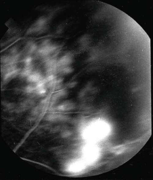
| A 44-year-old male patient with Eales disease developed migrainous headache and fatigue several years later. The patient presented with visual deterioration in the left eye in 1987. Ophtalmoscopy and fundus fluorescein angiography showed inflammatory lesions around the retinal blood vessels (Figure 1). Examinations for other causes of phlebitis were negative. Eales disease had been diagnosed. The patient was treated with photocoagulation and steroids. Six months later, he developed retinal detachment. He lost his vision after the surgical treatment. At the time of presentation, the patient had no neurological signs. The patient was admitted to our hospital for severe headache and fatigue. On arrival, he was alert and had no neck stiffness. Blood pressure was 120/80mmHg; Visual acuity was 20/20 in the OD and 0/20 in the OS. |
| |
|
|
Figure 1: Inflammatory lesions around the retinal blood vessels on fundus fluorescein Angiography. |
|
| |
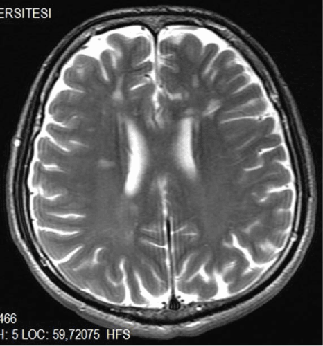
| Blood count, renal, hepatic profiles, PT (Prothrombin Time), PTT (Partial Thromboplastin Time), Erythrocyte Sedimentation Rate (ESR), C-Reactive Protein (CRP), protein-C,protein-S, antithrombin- III, vitamin A, E, B12 and Cerebrospinal Fluid (CSF) examination were normal. Rheumatoid factor, mutation of factor V Leiden, prothrombin 20210-A, and Methylenetetrahydrofolate Reductase (MTHFR), Antineutrophil Cytoplasmic Antibody (ANCA), Anti-nuclear antibody (ANA), antibodies of anticardiolipin and antiphospolipid were negative. Serology for viral hepatitits B and C were negative. Chest roentgenogram was normal. Brain stem auditory and somatosensory evoked potentials were normal. Left Visual Evoked potential was absent. Brain MRI showed multifocal white matter abnormalities (Figure 2). |
| |
|
|
Figure 2: Brain MRI, T-2 weighted axial image demonstrated multifocal white matter Lesions. |
|
| |
| Symptomatic treatment was initiated for migrainous headache and fatigue. Our patient responded well to the treatment of headache and fatigue. The patient’s control brain MRI showed no change at the end of one year. |
| |
| Discussion |
| |
| Eales disease is an idiopathic inflammatory venous occlusion that primarily affects the peripheral retina of adults and rarely affects CNS. The association of MS, myleopathy, spastic paraparesis, stroke and myelitis with Eales disease has been reported [3-6,11]. Vision loss is characteristically caused by bilateral, recurrent, vitreus hemorrhage and its sequelae. The etiopathogenesis of Eales disease is controversial and recognized as primary vasculitis of unknown etiology in young adults. Immune mediated mechanism has been proposed as a possible cause of Eales disease [1,2]. A recent report showed the serum and intraocular levels of MMP-9 and tumor necrosis factor-α (TNFα) in patients with Eales disease were increased. It seems to provide a plausible explanation for inflammation-mediated angiogenesis during the development of this condition [11]. Other studies suggest that certain peptide growth factors, for example platelet derived growth factor (PDGF), insulin-like growth factor I (IGF-I), epidermal growth factor (EGF), transforming growth factors (TGF-a and TGF-b), and vascular endothelial growth factor (VEGF), have a key role in the process of neovascularization by both direct and indirect means [12,13]. |
| |
| In earlier reports, neurological involvement in Eales Disease has been reported several times. Cerebral vasculitis, stroke, multiple sclerosis, migrainous headaches were described [5,7,14]. The value of white matter abnormalities in Eales disease without clinical findings hasn’t been defined. Our patient had migrainous headache and there were multiple white matter lesions in brain MRI. Also he didn’t have any neurological deficit and there was no attack in the past. The significance of white matter lesions seems to be unclear. But the probability of occurrence of a demyelinating disease like MS in the future should be kept in mind and neurological evaluation should be done intermittently. |
| |
| |
| References |
| |
- Biswas J, Sharma T, Gopal L, Madhavan HN, Sulochana KN, et al. (2002) Eales disease--an update. Surv Ophthalmol 47: 197-214.
- Das T, Pathengay A, Hussain N, Biswas J (2010) Eales' disease: diagnosis and management. Eye (Lond) 24: 472-482.
- Rodier G, Derouiche F, Bronner P, Cohen E (1999) [Eales disease with neurologic manifestation: differential diagnosis of multiple sclerosis. Report of two cases]. Presse Med 28: 1692-1694.
- Garg RK, Kar AM, Varma M (1993) Eales' disease with progressive spastic paraparesis. J Assoc Physicians India 41: 179.
- Opala G, Wajgt A, Ochudlo S, Marczewska E (1988) Eales disease and multiple sclerosis. Case report. Neurol Neurochir Pol 22: 340-342.
- Sawhney IM, Chopra JS, Bansal SK, Gupta AK (1986) Eales' disease with myelopathy. Clin Neurol Neurosurg 88: 213-215.
- Gordon MF, Coyle PK, Golub B (1988) Eales' disease presenting as stroke in the young adult. Ann Neurol 24: 264-266.
- Antigüedad A, Zarranz JJ (1994) Eales' disease involving central nervous system white matter. Neurologia 9: 307-310.
- Katz B, Wheeler D, Weinreb RN, Swenson MR (1991) Eales' disease with central nervous system infarction. Ann Ophthalmol 23: 460-463.
- Lejarreta-Andres S, Aguirregomozcorta-Gil M, Jimenez-Nieto M, Castellanos-Rodrigo M, Serena-Leal J (2010) [Recurrent stroke and myelitis in a patient with Eales' syndrome]. Rev Neurol 51: 445-447.
- Lejarreta-Andres S, Aguirregomozcorta-Gil M, Jimenez-Nieto M, Castellanos-Rodrigo M, Serena-Leal J (2010) Recurrent stroke and myelitis in a patient with Eales' syndrome. Rev Neurol 51: 445-447.
- Perentes Y, Chan CC, Bovey E, Uffer S, Herbort CP (2002) Massive vascular endothelium growth factor (VEGF) expression in Eales' disease. Klin Monbl Augenheilkd 219: 311-314.
- Angayarkanni N, Selvi R, Pukhraj R, Biswas J, Bhavesh SJ, et al. (2009) Ratio of the vitreous vascular endothelial growth factor and pigment epithelial-derived factor in Eales disease. J Ocul Biol Dis Infor 2: 20-28.
- Biswas J, Raghavendran R, Pinakin G, Arjundas D (2001) Presumed Eales' disease with neurologic involvement: report of three cases. Retina 21: 141-145.
|
| |
| |