| Research Article |
Open Access |
|
| Atteyat Selim* and Eman El-Nahas |
| Zoology Department, Faculty of Science, Tanta University, Tanta, Egypt |
| *Corresponding authors: |
Atteyat Seli
Department of Zoology, Faculty of Science
Tanta university, Tanta, Egypt
E-mail: Atteyat2000@hotmail.com |
|
| |
| Received February 07, 2012; Published July 27, 2012 |
| |
| Citation: Selim A, El-Nahas E (2012) Characterization of the Pars Distalis of the Egyptian Insectivorous Bats Taphozous Nudiventris by Using Both Ultrastructure and Histological Structure. 1: 212. doi:10.4172/scientificreports.212 |
| |
| Copyright:© 2012 Selim A, et al. This is an open-access article distributed under the terms of the Creative Commons Attribution License, which permits unrestricted use, distribution, and reproduction in any medium, provided the original author and source are credited. |
| |
| Abstract |
| |
| The ultrastructure and the histological structure of the pars distalis of the other species of bats in the world has been discussed, but there is little information available about the ultrastructure and histological investigations on the Egyptian bats, so the present investigation focus on the Egyptian insectivorous bats, in order to compare between the Egyptian bats and the others bats in the world. |
| |
| The material and methods: We collect the adult male animals from the field and caves, then decapitation and the pituitary glands put out. Some glands fixed in 10% formalin for histological observation and others fixed in glutaraldehyde for electron microscope. |
| |
| The results: The present investigation demonstrates that the gland is semicircular in shape, dorso ventrally compressed and there are different cell types in the pars distalis. STH cells are round to oval with eccentrically placed nucleus .The nucleus has different shapes, and there are numerous large size secretory granules but less number mitochondria in the cytoplasm in contract with the other bats in the world. LTH cells are characterized by large secretary granules than STH cells, dilated endoplasmic reticulum. ACTH cells present in groups with small granules collected at the periphery of the cells. The TSH cells are characterized by large and small size of secretory granules in contract with the other bats in world. |
| |
| The conclusion: Although all the species of bats were from the same phylum chiroptera , but the different in phylogeny and the different in habitat may be affected internally in the structure of pituitary gland tissue. |
| |
| Keywords |
| |
| Bats; Pars distalis; Ultrastructure; Histological structure |
| |
| Abbrevation |
| |
| N- Nucleus; S.G- Secretory Granules; M- Mitochonria; E.R- Enoplasmic reticulum; B.m- Blasma membrane; V- Vacules; |
| |
| Introduction |
| |
| In Egypt the insectivorous bats are monoestrous cycle from March to the end of July. We collect the specimens during this period .The ultrastructural study of the pars distalis has been mainly investigated, with the goal of defining not only microscopic characteristics but also the physiological significance of different cell categories with respect to reproduction, such as, LH, FSH and prolactin are fundamental to reproductive physiology. These cells of pars distalis not only play a role in reproductive processes of mammals but also changing in morphology during different phases of reproductive cycle of bats [1-6]. The ultrastructural and the functional characteristics of the anterior pituitary cells in the Indian fruit bat Rousettus Leschenaulti were studied by Bhiwgade, et al. [7]. In Myotis myotis the ultrastructural changes in gondatrophic and prolactin cells have been studied by Muniz, et al. [8]. In Vespertilionid bat, Scotophilus heathi electron microscopic studies revealed six distinct cell types in the pars distalis on basis of specific morphological characters, staining reactions and immunocharacteristics [9,10]. The ultra structural characterization of the pars distalis of the Indian female sheath-tailed bat, Taphozous longimanus by Archana and Mohan [11]. The light microscopic study of anterior pituitary reveals the presence of six cell types based on granulation and staining reaction in Myotis lucifugus lucifugus [12,13]. The cytological changes in the pituitary in relation to the sex-cycle in bats have been studied mostly in hibernating bats vespertilionids [13,14]. Six cell types have been recognized in the anterior pituitary on the basis of their morphological characteristics of the bat [15]. |
| |
| Materials and Methods |
| |
| For the present study the animals of Taphozous nudiventris were brought alive from Abo-Rawach –Giza –Cairo-Egypt in the laboratory. The specimens were killed by decapitation and the brain along with the pituitary was immediately out and fix for 24 h in 10% formalin for histological structure, dehydrated in a graded series of ethanol (70%- 100%), then cleared in xylol and embedded in paraffin wax. Serial sections of 6μm thick were obtained and stained with hematoxylin and eosin, Azan stain and PAS stain. For Transmission Electron Microscope (TEM) Romeis [16], the pituitary glands were fixed in fresh ice cold 3% glutaraldehyde for three hours and then four hours in 0.1 M cacodylate – buffered. The tissues were washed in buffer and then post fixed for one to two hours in 1% 0.067 M cacodylate- buffered osmium tetroxide. After dehydration with graded series of alcohol, the tissues were cleared in propylene oxide solution and embedded in araldite resin which would be polymerized at 60°C. Then, ultrathin sections from selected blocks were cut glass knife and picked up on 400-mesh copper gride. Sections were stained with toludine blue stain for semi thin, thin sections were examined under a JEM jeol-100s electron microscope. |
| |
| Results |
| |
| Histological observations |
| |
| In Taphozous nudiventris the gland is dorso ventrally compressed, and semicircular in shape. The anterior pituitary separated from the posterior by residual lumen while the pars intermedia is in close contact with the posterior pituitary (Figure 1). The cells of the anterior pituitary are distributed singly or small groups with the sinusoids .Two types of acidophilic cells and two types of basophilic cells were demonstrated by using azan stain and PAS reaction (Figure 2 and 3).Classification of the cell types is based on the staining properties of the cytoplasmic granules, their shape and size of the cells. |
| |
|
|
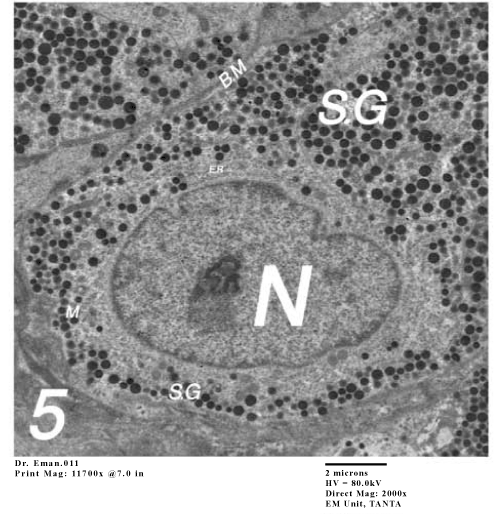
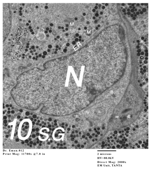
Figure 1: Electron micrograph of the pituitary gland of Tahozous nudiventris showing the STH cells. |
|
| |
|
|
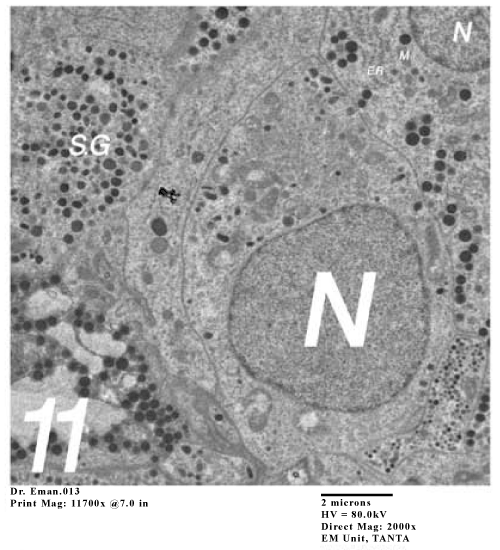
Figure 2: Electron micrograph of the pituitary gland of Tahozous nudiventris showing the STH cells. |
|
| |
|
|
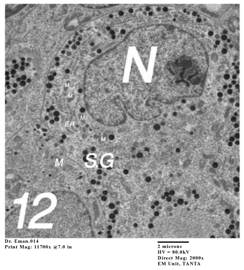
Figure 3: Electron micrograph of the pituitary gland of Tahozous nudiventris showing the STH cells. |
|
| |
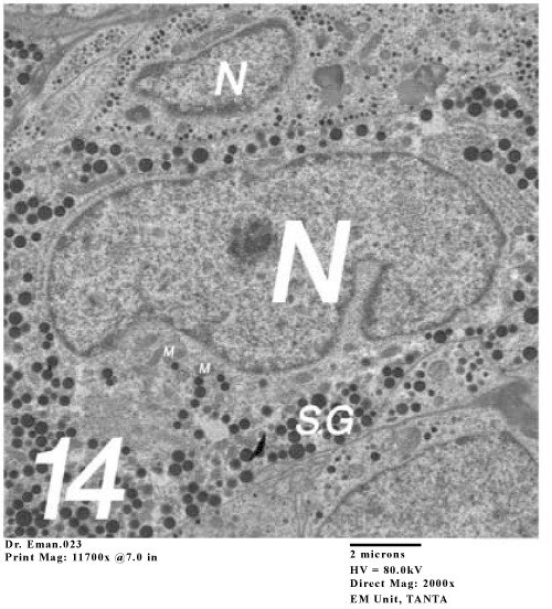
| Type-1 (probably STH cells): The secretory granules of these acidophilic cells are stained with acid dyes in various histochemical techniques used .Most of these cells are round in shape with spherical nucleus with mean diameter 5.33 ± 0.81mμm in both male and females (Figure 4). |
| |
| |
|
|
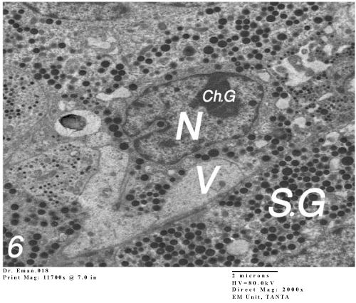
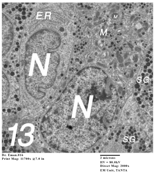
Figure 4: Electron micrograph of the pituitary gland of Taphozous nudiventris showing the LTC cells. |
|
| |
| Type-2 (Probably LTH Cells): These acidophilic cells are round but larger in size than the STH cell 6.21 ± 0.42mμm. It's distributed throughout the gland but is more towards posterior lateral and posteromedian regions of the anterior pituitary. By using PAS reaction, the LTH cells give more intensity red color than STH cells (Figure 5). |
| |
|
|
Figure 5: Electron micrograph of the pituitary gland of Taphozous nudiventris showing the LTC cells. |
|
| |
| The basophilic cells: The granulated cytoplasm of these cells react with the basic dyes of hematoxylin, aniline blue of azan stain but react very poor with PAS reaction (Figure 6 ) 4.25 ± 0.33mμm. |
| |
|
|
Figure 6: Electron micrograph of the pituitary gland of Taphozous nudiventris showing the LTC cells. |
|
| |
| The Electron microscope |
| |
| The current concept of cell identification and differentiation in the mammalian pituitary gland is based on the activity of the Golgiapparatus, the elaboration of secretary granules, liberation of the granules from the cell membrane, morphology of the mitochondria ,the shape and the size of the of the secretary granules and the cell nucleus. |
| |
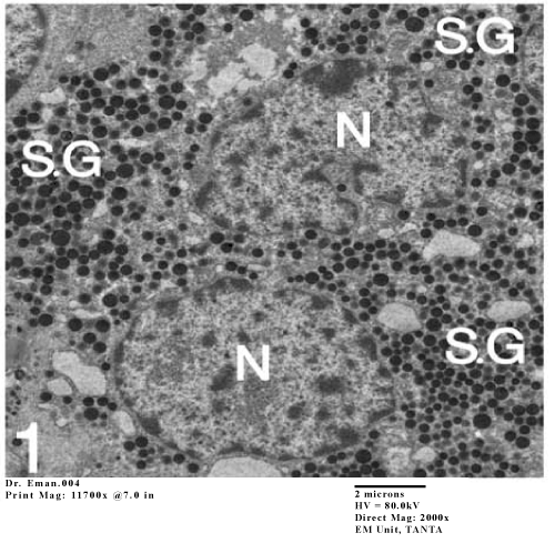
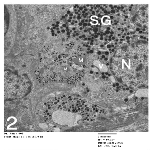
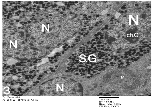
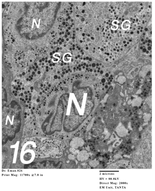
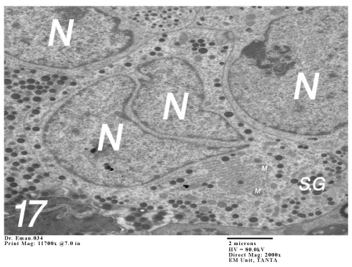
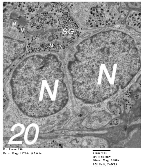
| STH-cell: These are the most numerous in the pars distalis of this bat. These are situated generally far from the capillaries .In the non pregnant females and the adult males, the nucleus is irregular in shape with dense chromatin granules and nucleolus is visible with a little indentation. The mitochondria is spherical, rough endoplasmic reticulum is not well developed and it is in the form of short tubular cisternae. Golgi complex is indistinct and there are numbers of large electron dense secretary granules (Figure 1 and 2). There is another STH cell which has lobed nucleus (Figure 3). |
| |
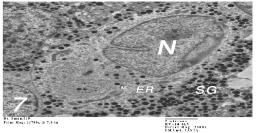
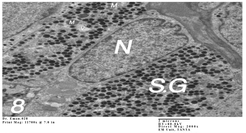
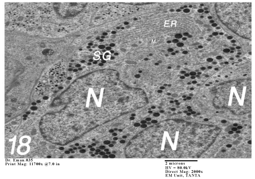
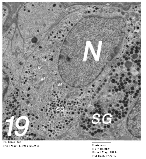
| LTH cells: LTH cells are easily recognizable with electron microscopy. The nucleus is eccentrically indented, the mitochondria is spherical or elongated, the endoplasmic reticulum is rough and the granules are more dense (Figure 4-8). |
| |
|
|
Figure 7: Electron micrograph of the pituitary gland of Taphozous nudiventris showing the LTC cells. |
|
| |
|
|
Figure 8: Electron micrograph of the pituitary gland of Taphozous nudiventris showing the LTC cells. |
|
| |
| Corticotroph (ACTH): These cells are found singly, irregular with eccentric nucleus. The nucleus is large, irregular with chromatin granules. Mitochondria are round, Golgi apparatus is not well developed, and Rough endoplasmic reticulum is in the form of elongated tubular profile arranged parallel to one another and is distributed throughout the cytoplasm. Secretary granules are spherical shaped. (Figure 19-21). |
| |
| Thyrotroph (TSH): These cells are found singly with centric nucleus. The nucleus is large with chromatin granules. Mitochondria are round, Golgi apparatus is not well developed, Rough endoplasmic reticulum present arranged parallel to one another and is distributed throughout the cytoplasm. Secretary granules are spherical and very small in size (Figure 9-13). |
| |
|
|
Figure 9: Electron micrograph of the gland Taphozous nudiventris showing the GTH cells and THC cells. |
|
| |
|
|
Figure 10: Electron micrograph of the gland Taphozous nudiventris showing the GTH cells and THC cells. |
|
| |
|
|
Figure 11: Electron micrograph of the gland Taphozous nudiventris showing the GTH cells and THC cells. |
|
| |
|
|
Figure 12: Electron micrograph of the gland Taphozous nudiventris showing the GTH cells and THC cells. |
|
| |
|
|
Figure 13: Electron micrograph of the gland Taphozous nudiventris showing the GTH cells and THC cells. |
|
| |
| Gonadotrophs (FSH and LH) Thyrotroph (TSH): These cells are found singly, angular in shape with eccentric nucleus. The nucleus is large, irregular with chromatin granules. Mitochondria are round, Golgi apparatus is not well developed, and Rough endoplasmic reticulum is in the form of elongated tubular profile arranged parallel to one another and is distributed throughout the cytoplasm. Secretary granules are spherical or ovoid shaped and exhibit variation in electron density (Figure 9 and 18). |
| |
|
|
Figure 14: Electron micrograph of the gland Taphozous nudiventris showing the GTH cells and THC cells. |
|
| |
|
|
Figure 15: Electron micrograph of the gland Taphozous nudiventris showing the GTH cells and THC cells. |
|
| |
|
|
Figure 16: Electron micrograph of the gland Taphozous nudiventris showing the GTH cells and THC cells. |
|
| |
|
|
Figure 17: Electron micrograph of the gland Taphozous nudiventris showing the GTH cells and THC cells. |
|
| |
|
|
Figure 18: Electron micrograph of the gland Taphozous nudiventris showing the GTH cells and THC cells. |
|
| |
|
|
Figure 19: Electron micrograph of the pituitary gland of taphozous nudiventris showing the ACTH cells. |
|
| |
|
|
Figure 20: Electron micrograph of the pituitary gland of taphozous nudiventris showing the ACTH cells. |
|
| |
|
|
Figure 21: Electron micrograph of the pituitary gland of taphozous nudiventris showing the ACTH cells. |
|
| |
| Discussion |
| |
| The morphology of the pituitary in Taphozous nudiventris is similar to that of the emballonurid bat, T. melanopogon [17,18]; rhinopomatid bat, Rinopoma hardwicki hardwicki Karim KB and Khan MS [19] and Megaderma lyra, Bhiwagade DA et al. [20]. The present study demonstrates that the different types of gonadotrophs and lactotrophs in the anterior pituitary in both sexes undergo changes in number and cytological characters parallel with the reproductive periodicity. |
| |
| The acidophilic cells, due to their solid protein nature of high insolubility, are easily preserved by any method of fixation. Type I cells are observed in the pituitary of mature males and females. In the present study, orange color STH and red color LTH cells can be differentiated by PAS reaction techniques are present few in number in non pregnant females and their number increases similar to that reported in T. melanopogon and Cynopterus sphinx [3]. In females apart from the lactogenic function of the LTH cells they are also found to be luteotrophic in function. In the hibernating bat, Miniop/erus schreibersii which shows delayed implantation, the LTH cells are observed at the time of implantation and activation of the corpus luteum after hiberoation [21]. |
| |
| The present study shows the basophiles with the azan stain and PAS reaction. In H. speoris, LH cells can be distinguished when the corpus luteum is regressing following the formation of choriovitelline placenta. These cells then increase in size and number, and subsequently involute at the termination of pregnancy and lactation. In C. sphinx, during the first pregnancy cycle, a few cells are visible at estrus and ovulation and remain so until mid pregnancy. At late pregnancy, no LH cells are visible. In T kachhensis, large numbers of FSH cells are observed in the anterior pituitary of estrus female. |
| |
| Electron microscopic study reveals the state of activity of the cells and their morphological features in the non pregnant adult females and adult males, which allows formulating the criteria for their functional analysis. In conjunction with our knowledge of the ultrastructure of the mammalian pituitary gland Herlant [22], Harris & Donovan [23], Nunez et al. [24], Bhiwgade et al. [7], Muniz et al. [8], Singh & Krishna [9], the ultrastructural observations demonstrate the presence of six cell types in the pars distalis of the bat Taphozous nudiventris. |
| |
| The increase in nuclear size, enlargement of nucleoli and nature of secretary granules of the cells has taken as the indicators of differentiation between the different cells. |
| |
| Somatotroph (STH- cell): Ultrastructural characteristics of STH cells of Indian fruit bat, R. leschenaulti have been studied by Bhiwgade et al. [7]. These cells are round to oval in shape with centrally placed round nucleus. The secretary granules are numerous, uniform, round and very dense about 350-400 nm in diameter. The Golgi apparatus is inconspicuous and mitochondria are round and scattered in the cytoplasm. The STH cells in the present bat Taphozous nudiventris are characterized by different shapes of the nucleus from rounded, irregular to bilobed. The secretary granules are numerous and large, no well developed Golgi apparatus, but in S. heathi, Singh and Krishna [9], the secretary granules are dense ranging from 240 to 480nm in diameter, round mitochondria and Golgi apparatus present in the STH cells. |
| |
| In Hipposideros lankadiva, Seraphim ER, [25] STH cells are round to oval with eccentrically placed nucleus. The secretary granules are numerous, mostly round to oval with uniform electron density. The well developed Golgi, endoplasmic reticulum and small amount of secretary granules indicate a cell under vigorous synthetic activity while those filled with secretary granules with reduced Golgi complex suggest reserve or storage state of cells, supporting the present observations of the non pregnant female. |
| |
| The Lactotrophic Cells (LTH cells): The ultrastructure features of LTH cells in the pars distalis of non pregnant bats differ from that of the other cells of the estrus bat. Mitochondria are numerous, rough endoplasmic reticulum are well developed and the electron dense granules are very large and are seen scattered in cytoplasm. The size of granules ranges from 300-500 nm diameter. |
| |
| Bhiwgade et al. [7] reported that the LTH cells are characterized by the largest secretary granules, well developed endoplasmic reticulum and numerous mitochondria in the cytoplasm. They further reported that an accumulation of secretary material in the PRL cells associated with the retention of milk following the removal of litter contributes favorably for identification of the prolactin in bat. An accumulation of secretary granules in PRL cells after 8 hrs of non suckling and decrease of granules prompted by 10 minutes of suckling has been confirmed, confirming to the present study. |
| |
| In S. heathi, Singh and Krishna [9] reported numerous mitochondria, dilated endoplasmic reticulum and extensive Golgi complex, large number of secretary granules in the LTH cells. Ishibashi T and Shiino M [26] identified the LTH cells in the pars distalis of Pipistrellus abramus. These cells show morphologically active features in pregnant and lactating females and have a tendency to contain smaller secretary granules. |
| |
| The proliferation activity of mammotrophs in female bat was observed during pregnancy and lactation [27]. The morphometrical activity and PRL level were mostly low during follicular development and the early implantation and this activity and PRL level increases after implantation [28]. |
| |
| The ultrastructural observations on LTH cells showed that size of secretary granules vary from species to species Richardson BA [4] The size of secretary granules in bat, M. myotis ranges from 170-550 nm diameter Herlant [29] R. leschenaulti from 400-600 nm diameter; S. heathi, from 300-600 nm diameter [9] . In the present study the size of secretary granules in Taphozous nudiventris ranges from 300 to 500 nm diameters. The LTH cells, thus identified in pars distalis of non pregnant and pregnant bat with electron microscopy are comparable with LTH cells of the other species of bats. |
| |
| In the present study, LTH cells in par distalis of non pregnant bat, which is a continuous breeder, are synthetically active and elaborate large quantity of prolactin during pregnancy. This hormone may be stimulating luteal cells of corpus luteum to synthesize progesterone. The ultrastructural features of luteal cells in Taphozous indicate that these cells are steroidogenically active during pregnancy [30], suggesting that the LTH cells are luteotrophic in this species of bat as observed in other bat species. Our findings correlate with the findings of other workers on bats. |
| |
| Corticotroph (ACTH cells): In the present study the ACTH cells are elongated or angular found either singly or in groups in pars distalis. Mitochondria are round with lamellar cristae. Rough endoplasmic reticulum is in the form of elongated and short tubular profiles. Golgi complex is inconspicuous. The spherical or ovoidal secretory granules (200-250 nm in diameter) of variable electron density are present just below the plasma membrane, which similar to the case of the ACTH cell of T. longimanus. |
| |
| Similar ultrastructural characteristics of ACTH cells are reported also in S. heathi [9], R. leschenaulti [7], H. lankadiva [25]. |
| |
| Thyrotroph (TSH cells): The TSH cells show ultrastructural features that observed in the TSH cells during estrus, except well developed rough surfaced endoplasmic reticulum. It is in the form of array of elongated, tubular or lamellar cisternae frequently localized at one pole or periphery of the cell. The profiles of cisternae are parallel to one another or curved. Golgi is inconspicuous. The small secretary granules are very small 150-200 nm in diameter, in varying electron density are scattered throughout the cytoplasm or show peripheral distribution. Thus, ultrastructural features of TSH cells in the non pregnancy suggest that these cells elaborate large quantity of thyrotrophic hormones. In S. heolhi, TSH cells showed the presence of a few secretary granules which ranged in diameter from 100-160 nm. |
| |
| Gonadotrophs (FSH and LH cells): Several researchers have shown by electron microscopy [31,32] that morphological differences between FSH and LH producing cells exist. Herlant [1,22,29], Azzali [18] and Bhiwgade et a1. [7] studied cytological variations in gonadotrophs in different species of bats under various physiological conditions. They have differentiated the gonadotrophs into two distinct entities, the FSH and LH secreting gonadotrophs. According to Bhiwgade et al. [7] the variations observed in the electron density of the secretory granules is sufficient to differentiate two types of gonadotrophs. These cells also differ in their cytoplasmic organelle. The present ultrastrutctural studies on par distal are from non pregnant bats, Taphozous nudiventris point to the existence of two types of gonadotrophs in bats. The FSHsecreting cells described in bats correspond to the presumptive rat FSH cells of Nakane [33] bat FSH cells of Herlant [22] and mink FSH cells of Murphy & James. The LH gonadotrophs in bats correspond to the LH cells of Nakane, gonadotrophs of Rennels el at. and LH cells of Herlant [22]. |
| |
| FSH cells in the pars distalis of non pregnant female are large ovoid and the rough surfaced endoplasmic reticulum is well developed. Secretary granules are spherical, 200-400 nm in diameter and show variable electron density, nucleus is irregular in outline. Large number of mitochondria with collapsed cristae is present in the cytoplasm. The dilated cisternae of rough endoplasmic reticulum are sparsely dotted with ribosome and are distributed throughout the cytoplasm. Thus the cytoplasm appears vacuolated. |
| |
| In the present study, the cell types, FSH, LH and LTH in the pars distalis of T. longimanus during pregnancy are active, elaborating hormones which are needed for the development of embryo in one uterine horn and folliculogenesis in contralateral horn as this bat is continuous breeder and pregnancies follow in quick succession. As this bat is continuous breeder and pregnancies follow in quick succession. |
| |
| Acknowledgement |
| |
| The first thanks is to Allah. The electron microscopy facilities provided by Dr. Azat, Department of Histology, Faculty of medicine for their excellent technical assistance. Our thanks are due to also to Professor, Dr. Gamal Madkour, who introduces to me all facilities of bat in our Department. |
| |
| |
| References |
| |
- Herlant M (1962) Electron microscope studies on the adenohypophysis of Myotis myotis during gestation. Gen Compo Endocr 2: 631.
- Peyre A, Herlant M (1963) Correlations hypo-physo-gcnitales chez la femelle du Minioptere (Miniopterus schreibersii B). J Compo Endocrinol 3: 726-727.
- Badwaik NK (1988) Cytology and seasonal changes of the pituitary of the emballonurid bat. Taphozous melanopogon (Temnick) Proc Indian Acad Sci (Anim Sci); 97: 479-89.
- Richardson BA (1979) The anterior pituitary and reproduction in bats. J Reprod Fertil; 56: 379-389.
- O'Brien GM (1996) Comparative morphology of the pituitary gland in Australian flying foxes (Megachiroptera: genus Pteropus). Anat Rec; 244: 70-77.
- O'Brien GM, McFarlane 1R, Kearney PJ (2003) Pituitary content of luteinizing hormone reveals species differences in the reproductive synchrony between males and females in Australian flaying- foxes (genus Pteropus). Reprod Fertil Dev; 15: 255-261.
- Bhiwgade DA, Akolkar YY, Menon SN, Manekar AP, Senad DG (1989) Ultrastructural and functional characteristics of anterior pituitary cells in the Indian fruit bat, Rousettus leschenaulti (Desmarest). Acta Anat (Basel); 135:129-141.
- Muniz E, Jimenez L, Gragera R, Fernandez A, Rua C (1991) Ultrastructural changes in the gonadotrophic and prolactin cells of Myotis myotis under experimental conditions. Funet Dev Morphol; 1: 15-18.
- Singh UP, Krishna (1994) Identification, localization and distribution of different cell types in adenohypophysis of female Vespertilionid bat, Scotophilus heathi: A combined histochemical, immunocytochemical and electron microscopic study. Proc Insl Nat Sci Acad 60: 115-127.
- Singh UP and Krishna A (1997) Pituitary Adrenocorticotrophic (ACTH) cells during reproductive cycle in a Vespertilionid bat, Scotophilus heathi. Acta BioI. Hung 48: 409-420.
- Archana A, Mohan M (2010) Ultrastructural Characterization Of The Pars Distalis Of The Indian Female Sheath-tailed bat, Taphozous longimans. Int J Morphol 28: 787-801.
- Gretchen LH (1979) Animal tissue techniques. (4th edn) Philadelphia, WH Freeman Company.
- Siegel JH (1955) J Morph 96: 223-263.
- Herlant M (1968) Cycle sexuel chez les chiropteres des regions temperees. 1n: Cycles Genitaux Saisonniers de Mammiferes, Canivene, R (ed) Paris, Masson et Cle pp: 1ll-131.
- Nerkar AA, Gadegone MM (2011) Seasonal variations in anterior pituitary cells types of the emaballonurid bat, Taphozous kachhensis. Journal of cell and tissue research 11: 12507-2513.
- Romeis B (1989) Mikroskopische Technik.Urban and chwarzenberg , Munchen wien Bultimore; 30: 144-1990.
- Azzali G (1971) Citologie adenoipofisaria dei Chirotteri con particolare riguardo aile cellule FSH, LH, ACTH e LTH. Ateneo Parmense Sezione I, Acta Biomed 42: 169-229.
- Azzali G (1971) Ateno parmence. Acta Bio-Mcd 42: 92-29.
- Karim KB, Khan MS (1985) J Curro Bio Sci 2: 6168.
- Bhiwagade DA, Sapkal YM, Manekar AG (1982) J Bombay Nat Hist Soc 78: 103-117.
- Peyre A, Herlant MCR (1963) hbd Seanc Acad Sci, PansD 257: 524-526.
- Herlant M (1964) The cells of the adenohypophysis and their functional significance. 1nt Rev Cytol; 17: 299-382
- Harris GW, Donovan BT (1966) The Pituitary gland. London, Butterworths, pp.586, 670.
- Nunez EA, Gershon M D (1980) Phasic secretion by follicular cells of the bat adenohypophysis during the prearollsal period of their annual life cycle. Am 1 Anal; 165: 111-120.
- Seraphim ER (2004) Endocrine interaction during different phases of the female reproductive cycle in Hipposideros lankadiva (Kelaart). Ph.D. thesis, RTM Nagpur Uni, Nagpur, India.
- Ishibashi T, Shiino M (1989) Subcellular localization of prolactin in the anterior pituitary cells of the female Japanese house bat, Pipistrellus abramus. Endocrinology; 124: 1056-1063.
- Kawamoto K (2003) Endocrine control of the reproductive activity in hibernating bats. Zool Sci; 20: 1057-1069.
- Anthony, ELP (2000) Endocrinology of reproduction in bats: central Control. In: Crichton, E G & Krutzsch, PH Reproductive Biology of Bats. London, Academic Press, pp: 1-26.
- Herlant M (1963) Apport de la microscopie electronique a I' etude du lobe anterieur de l'hypophyse. 1n: Cytologie de I' Adenohypophyse. Benoit, 1 & Dalage, C (Eds) Paris, Editions du Centre National dela Recherche Scientifique, pp: 73-90.
- Nerkar AA, Gadegone MM (2007) Ultrastructural organization of the corpus luteum of the Indian emballonurid bat, Taphozous longimanus (Hardwicke). 1 Endocrino Reprod; 2: 76-81.
- Farquhar MG, Rinehart JF (1954) Electron microscopic studies of the anterior pituitary gland of castrate rats. Endocrinology; 54: 516-541.
- Purves HD (1661) Morphology of the hypophysis related to its function. In: Young W C (ed) Sex and Internal Secretion, London, B & Cox, T Ltd pp: 162-229.
- Nakane PK (1970) Classification of anterior pituitary cell types with immunoenzyme histochemistry. 1 Histochem Cytochem; 18: 9-20.
|
| |
| |