| Research Article |
Open Access |
|
| Muhammad Aliyu*1,2, Oyeronke A Odunola2, Solomon E Owumi2, Nathan Habila1, Idowu A Aimola1 and Ochuko L Erukainure3 |
| 1Department of Biochemistry, Ahmadu Bello University, Zaria, Nigeria |
| 2Department of Biochemistry, University of Ibadan, Ibadan, Nigeria |
| 3Food Technology Division, Federal Institute of Industrial Research, Oshodi, Nigeria |
| *Corresponding authors: |
Muhammad Aliyu
Department of Biochemistry
Ahmadu Bello University
Zaria, Nigeria
Tel: +2347038161430
E-mail: amachida31@gmail.com |
|
| |
| Received July 07, 2012; Published August 07, 2012 |
| |
| Citation: Aliyu M, Odunola OA, Owumi SE, Habila N, Aimola IA, et al. (2012) Ethanol Suppresses the Effects of Sodium Arsenite in Male Wister Albino Rats. 1: 222. doi:10.4172/scientificreports.222 |
| |
| Copyright: © 2012 Aliyu M, et al. This is an open-access article distributed under the terms of the Creative Commons Attribution License, which permits unrestricted use, distribution, and reproduction in any medium, provided the original author and source are credited. |
| |
| Abstract |
| |
| Background: Millions of people around the world get exposed to high levels of heavy metals in drinking-water. Therefore, quality control in drinking-water, food industries and detection of heavy metals is an extremely critical issue in maintaining the human health. Of such heavy metals is arsenic. Water as one of the major ingredient in both traditional and modern beer fermentation process may/may not be contaminated with arsenic due to poor quality control. As a result people are exposed to the end product which constituted arsenic compound. Sodium arsenite and ethanol has been established to be toxic both in vivo and in vitro. |
| |
| Objective: This study is primarily designed to investigate the effect of co-administration of sodium arsenite and ethanol in male Wister albino rats. |
| |
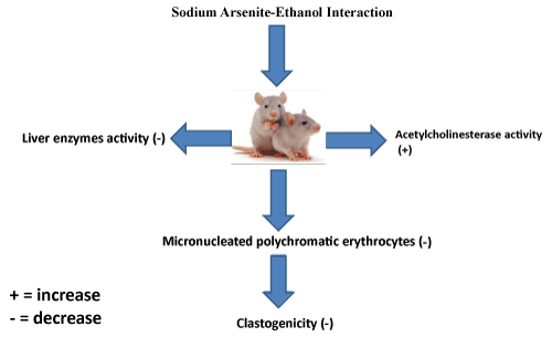
| Methods: Forty five (45) albino rats divided into nine (9) groups of five rats each were administered with distilled water, 2.5mg and 5mg/kg body weight of sodium arsenite, 3% and 6% (v/v) respectively. Treatment was based on single and combined administration. Micronucleated polychromatic erythrocytes, acetylcholine esterase, Aspartate Transaminase (AST), Alkaline Phosphatase (ALP), Alanine Transaminase (ALT) and hematological parameters were determined by standard procedures (Figure 1). |
| |
| Results: Combined treatment with sodium arsenite and ethanol significantly decreased the number of micro nucleated polychromatic erythrocytes, AST, ALT and ALP activities as against the single treated groups. However, there was stabilization of acetylcholine esterase activity in the brain. The hematological parameters level was also stabilized. |
| |
| Conclusions: We therefore proposed that the chemical interaction between sodium arsenite and ethanol is what was responsible for the suppression of sodium arsenite – induced clastogenic, hepatotoxic, anti-acetylcholine esterase and anemic effects. |
| |
| Keywords |
| |
| Sodium arsenite; Ethanol; Micronucleated polychromatic erythrocyte; Acetylcholine esterase, Hematology |
| |
| Introduction |
| |
| Continuous exposure of humans to arsenic through long-term ingestion of contaminated water and its attendant health problems has been widely reported. Inorganic arsenic compounds are widely distributed in nature and a lot of epidemiological evidence exist associating occupational and environmental exposure to them with human carcinogenesis [1,2]. For instance, exposure to trivalent and pentavalent forms of arsenic, which occurs worldwide primarily through occupational and environmental exposure, causes characteristic skin alterations (ulceration), including hyperkeratosis and skin cancer [3]. Epidemiological studies conducted in Taiwan [4], Chile [5] and Japan [6] indicated a connection between arsenic exposures from contaminated drinking water among the inhabitants. It is also known that arsenic interact with other substances, metals inclusive there by potentiating its effects and/or vice versa [7]. There is growing evidences that sodium arsenite intoxication can compromise the integrity of the liver in mouse, rat, fish, and goat [8,9,10]. Recently, some studies suggest the use of antioxidants and antioxidant rich foods and herbal medicinal plant for the management of arsenicosis [11]. Induction of cancer is frequently associated with DNA damage, changes in ploidy of cells, and non-random chromosome aberrations which can result from exposure to arsenic [12]. Association between chronic alcohol abuse and the development of cirrhosis, as well as between cirrhosis and the development of Hepatocellular Carcinoma (HCC), is well documented [13,14]. Acute and chronic ethanol treatment has been shown to increase the production of reactive oxygen species, lower cellular antioxidant levels, and enhance oxidative stress in many tissues, especially the liver [15]. Ethanol-induced oxidative stress plays a major role in the mechanisms by which ethanol produces liver injury [15]. Water as one of the major ingredient in both traditional and modern beer fermentation process may/may not be contaminated with arsenic due to poor quality control. As a result people are exposed to the end product which constituted arsenic compound. Sodium arsenite and ethanol has been established to be toxic both in vivo and in vitro. Therefore, this study was undertaken to investigate the effect of coadministration of sodium arsenite and ethanol on male Wister albino rats with a view to establish the facts on whether or not their interaction could be enhance or suppress by one another. |
| |
| Materials and Methods |
| |
| Chemicals |
| |
| Sodium Arsenite (NaAsO2 , Mol.wt 129.91, 98% prod 30110, BDH chemicals Ltd Poole England) and ethanol (C2H5OH, Mol.wt 46.07, 99.7 to 100% v/v pod 10107, BDH chemicals Ltd Poole England) were dissolved in distilled water to prepare 3%, and 6% ethanol (v/v). |
| |
| Animals |
| |
| Forty five (45) male albino rats weighing 151 – 198 g were used for the present investigation. They were reared at the animal house of the Department of Pharmacy, Faculty of Pharmaceutical Sciences, Ahmadu Bello University, and Zaria, Nigeria with the approval of animal rights review committee. They were acclimatized for 1 week on normal diet of pelletized mouse chow, with water given ad libitum at room temperature within a 12-h light and dark cycle before the commencement of the experiment. |
| |
| Experimental design |
| |
| The animals were divided into nine different groups of five (5) rats each according to their body weight proximity and treated once in a week for a period of four week as shown below: |
| |
| Group 1: Distilled water |
| |
| Group 2: 2.5mg/kg body weight of sodium arsenite |
| |
| Group 3: 5mg/kg body weight Sodium arsenite |
| |
| Group 4: 3% ethanol (v/v) |
| |
| Group 5: 6% ethanol (v/v) |
| |
| Group 6: 2.5mg/kg body weight Sodium arsenite + 3% ethanol (v/v) |
| |
| Group 7: 5mg/kg body weight Sodium arsenite + 6% ethanol (v/v) |
| |
| Group 8: 2.5mg/kg body weight Sodium arsenite using 3% Ethanol as solvent (v/v) |
| |
| Group 9: 5mg/kg body weight Sodium arsenite using 6% Ethanol as solvent (v/v) |
| |
| Twenty-four (24) hours after the last administration, animals were sacrificed by using 60mg/kg body weight of sodium pentothal. Liver, Brain, Femur and Blood samples were collected, part of the blood samples were placed in Ethylene Diamine Tetra acetic Acid (EDTA) bottles for hematological analysis and the remaining were centrifuged at 4,000 rpm for 5 minutes to obtain the serum for analyses. The liver and brain weight were immediately taken and brain was quickly placed on an inverted Petri dish on ice. The fore brain was dissected, weighed and homogenized in 10ml of a medium containing 10mM Tris – HCl buffer (pH 7.2) and 160mM sucrose. The total homogenate was centrifuged at 3500g at - 4°C in a refrigerated centrifuge for 5 minute. The supernatant was used for Acetylcholine Esterase (AChE) activity. The femurs were preserved for micronucleus assay. |
| |
| Determination of biochemical and hematological parameters |
| |
| The Alkaline Phosphatase (ALP), Alanine Aminotransferase (ALT), and Aspartate Aminotransferase (AST) were determined by using Auto Analyzer Hitachi Roche 7020 (902), Japan Inc. according to manufacturer’s protocols. Complete blood counts were also determined using Coulter HmX Hematology Analyzer Beckman Coulter Inc. according to manufacturer’s protocols. |
| |
| Determination of acetylcholine esterase activity |
| |
| The activity of AChE in the brain was determined by the method described by Ellman et al. [18], as modified by Srikumar et al. [19] The mixture was prepared by mixing 0.4mL aliquot of the homogenate and added to 2.6ml phosphate buffer (0.1M, pH8.0) and 100μL of DTNB (270μM). This was pre-incubated for 2 minute at 30oC and the reaction was started with the addition of 20μL ATC (30mM). The product of thiocholine reaction with DTNB was determined at 412nm for a period of 10 minute at 2 minute intervals. The absorbance per minute was determined. |
| |
| Micronucleus assay |
| |
| Clastogenic effects were evaluated in the rat bone marrow using the micronucleus assay as described by Heddle and Salmone, [20] and modified by Heddle, et al, [21]. Two hours prior to sacrifice, the animals were injected (i.p.) with 0.04 % colchicine (1ml/100g body weight). Bone marrow cells from both femurs were used for preparing slides. The slides were fixed, air-dried and pretreated with May-Grunewald solution. They were then stained with 5% Giemsa solution. The slides were scored for the presence of micronucleated polychromatic erythrocytes (mPCEs) in 1000 cells according to standard procedure. |
| |
| Statistical analysis |
| |
| The results were expressed as mean ± Standard error. Differences between the groups were analyzed by one-way analysis of variance (ANOVA) with the aid of Statistical Package for Social Sciences (SPSS) software, SPSS Inc., Chicago, Standard version 10.0.1. P-values < 0.05 were considered statistically significant for differences in mean using the Least of Significance Difference (Lsd). |
| |
| Results |
| |
| The body weight results from table 1, shows a significant (p<0.05) decrease in % weight change in a concentration dependent manner with concomitant significant (p<0.05) increase in the groups treated with sodium arsenite and ethanol together. From table 1 there are no significant (p>0.05) difference on the organs and relative organs weight. |
| |
|
|
Table 1: Body and Organ weight (g) for n=5, (mean ± SE) of the experimental animals before and after exposure to sodium arsenite and ethanol. |
|
| |
| Based on the activity of the liver enzymes (ALP, ALT and AST) from table 2, the interaction of ethanol and sodium arsenite significantly (p<0.05) decrease the activity of the liver enzymes as compared with the groups treated with sodium arsenite, ethanol alone. This might suggest the fact that ethanol is suppressing the effect of sodium arsenite. |
| |
|
|
Table 2: Serum level mean ± S.E (U/L) for n=5, of aspartate amino transferase, alanine amino transferase and alkaline phosphatase in sera of rats. |
|
| |
| As depicted in table 3, it was observed that administration of ethanol and sodium arsenite significantly (p<0.05) decreased the number of micronuclei/1000PCE as compared with the groups treated with sodium arsenite, ethanol respectively, indicating the fact that the ethanol administration suppressed the clastogenic effect of sodium arsenite. This might further suggest an interaction between sodium arsenite and ethanol which led to the observed anticlastogenic activity as sodium arsenite is a known clastogen. |
| |
| |
|
|
Table 3: Induction of micronucleated polychromatic erythrocytes (mPCEs) in rat bone marrow cells after exposure to sodium arsenite and ethanol mean ± SE for n=5. |
|
| |
| From table 4, the activity of acetylcholine esterase in the brain was significantly (p<0.05) inhibited by sodium arsenite in a concentration dependent manner. At low concentration ethanol did not significantly (p>0.05) inhibit the activity of the enzyme but did at a higher concentration. However, the co-administration of sodium arsenite and ethanol led to an increased activity of acetylcholine esterase suggesting that the interaction might have caused the enzyme induction. |
| |
|
|
Table 4: Acetylcholine esterase activity mean ± SE for n=5, in the brain of the rats after exposure to sodium arsenite and ethanol. |
|
| |
| However, from table 5, most of the haematological parameters PCV, Hb and RBC were significantly (p<0.05) decreased with increased sodium arsenite and ethanol concentration but these were observed to start increasing from group 6 to 9. Furthermore, in terms of WBC it was only group 8 that showed a significant (p<0.05) decrease while others showed no remarkable increase in WBC. This signifies that the simultaneous administration of the toxicants in question has a counteracting effect, thus suggesting that the interaction is possibly having anti-anaemic effect. |
| |
|
|
Table 5: Results of Heamatological parameters mean ± SE for n=5, of rats after exposure to sodium arsenite and ethanol. |
|
| |
| Discussion |
| |
| Exposure to arsenite has been linked to diverse defects in both experimental animals and in humans [22-26]. The liver is an important target organ for arsenic toxicity [27]. Arsenic has been claimed to be of clinical utility in the treatment of syphilis, amoebiasis, and certain other tropical diseases [24] and also has been used in Fowler solution in the treatment of arthritis [24], but recently arsenic intoxication in experimental animals has been associated with hepatic tumours [25], the inhibition of testicular steroid genic function [28], and spermatogenesis [26], as well as with severe metabolic disorders such as diabetes in humans [22,23]. It is known that (SA) can act as comutagen due to its ability to inhibit the activities of thiol containing enzymes [27], such as DNA ligase [28] resulting in defective DNA replication, repair, recombination and joining of single- and double-stranded DNA breaks [29]. |
| |
|
|
| |
| Ethanol-induced oxidative stress is as a result of the combined impairment of antioxidant defences and the production of reactive oxygen species by the mitochondrial electron transport chain, the alcohol-inducible Cytochrome P450 (CYP) 2E1 and activated phagocytes [15]. Furthermore, Hydroxyethyl Radicals (HERs) are also generated during ethanol metabolism by CYP2E1. The available evidence indicates that, by favouring mitochondrial permeability transition, oxidative stress promotes hepatocyte necrosis and/or apoptosis and is implicated in the alcohol-induced sensitization of hepatocytes to the pro-apoptotic action of TNF-a. Moreover, oxidative mechanisms can contribute to liver fibrosis, by triggering the release of pro-fibrotic cytokines and collagen gene expression in hepatic stellate cells [30]. |
| |
| This study examines the effect of co-exposure to sodium arsenite and ethanol on male Wister albino rats. The interaction between sodium arsenite and ethanol seem to reverse the effect of decreased body weight. This might be attributed to the fact that ethanol induces fatty liver with enhanced lipogenesis that ultimately lead to an increase in weight [31]. The results of the present study clearly demonstrate that administration of sodium arsenite and ethanol respectively, significantly (P < 0.05) induced the formation of micronuclei in the Polychromatic Erythrocytes (PCEs) of the rat bone marrow cells. However, the reversal of that happened when both toxicants were coadministration. This is may be due to the fact that arsenite generates free radicals that can attack DNA leading to chromosomal breakage. In addition, acetaldehyde the end product of ethanol metabolism can form DNA adducts which might also explain why groups treated with sodium arsenite and ethanol respectively were able to induced clastogenicity. The results obtained from the assessment of the serum activities of ALP, ALT and AST shows that their activities increased in a concentration dependent manner. Interestingly, the activities were decreased in the groups of co-administration providing a clue that the chemical interaction between sodium arsenite and ethanol is having a reversal effect. Exposure to sodium arsenite had been shown to induce ALP, AST and ALT activity [32], which is clearly an indication of induction of hepatotoxicity and oxidative stress in the hepatocytes. |
| |
| Acetylcholine (ACh) is a neurotransmitter that functions in conveying nerve impulses across synaptic clefts within the CNS [33]. Following the transmission of an impulse across the synapse by the release of Ach, AChE is released into the synaptic cleft [34]. This enzyme hydrolyzes ACh to choline and acetate and transmission of the nerve impulse is terminated [35]. The same scenario surfaced on acetylcholine esterase activity being suppressed as concentration of the toxicants increased and elevated in the co-administered groups; probably the interaction might have initiated an enzyme induction as the activity of AChE is vital to neurological functions. Similarly, based on the hematological parameters single and co-administered groups were depicting an antagonistic effect. This suggests the possibility of an interaction between sodium arsenite and ethanol to have a stabilizing effect on the levels of hematological parameters as the toxic compounds like CCl4, arsenic and ethanol has been found to negatively affect the levels of these parameters [36]. It is against this background we proposed that the reaction between sodium arsenite and ethanol in the presence of water generates dimethyl hydroxyl arsenous acid which ultimately make it more polar and as such easily excreted without necessarily causing harm to the system. |
| |
| Conclusion |
| |
| We therefore conclude that the chemical interaction between sodium arsenite and ethanol is what is responsible for the suppression of sodium arsenite – induced clastogenic, hepatotoxic, anti-acetylcholine esterase and anemic effects by ethanol. This might be reason why most people that are addicted to alcoholics don’t easily come down with arsenic poisoning even though they are exposed to contaminated drinking water. We recommend further molecular studies in this regard. |
| |
| Potential Conflicts of Interest |
| |
| There are no conflicts of interest. |
| |
| Acknowledgments |
| |
| We graciously appreciate the consistent laboratory effort of Azuibuke Okafor, edet imaikop, Fauziyya Abdullahi, Hope Ataboh, Victory E. and Malam Aliyu (laboratory technologist) all from the Department of Biochemistry, Ahmadu Bello University, Zaria, Nigeria. |
| |
| |
| References |
| |
- Loprieno N (1975) Letter: International Agency for Research on Cancer (IARC) monographs on the evaluation of carcinogenic risk of chemicals to man: "Relevance of data on mutagenicity". Mutat Res 31: 210.
- Arteel Gavin E, Luping G, Schlierf Thomas (1981) Inorganic arsenic compounds. J Chem Soc Perkin Trans 1: 2563.
- Yoshida T, Yamauchi H, Fan Sun G (2004) Chronic health effects in people exposed to arsenic via the drinking water: dose-response relationships in review. Toxicol Appl Pharmacol 198: 243-252.
- Chiou HY, Hsueh YM, Liaw KF, Horng SF, Chiang MH, et al. (1995) Incidence of internal cancers and ingested inorganic arsenic: a seven-year follow-up study in Taiwan. Cancer Res 55: 1296-1300.
- Smith AH, Goycolea M, Haque R, Biggs ML (1998) Marked increase in bladder and lung cancer mortality in a region of Northern Chile due to arsenic in drinking water. Am J Epidemiol 147: 660-669.
- Tsuda T, Babazono A, Yamamoto E, Kurumatani N, Mino Y, et al. (1995) Ingested arsenic and internal cancer: a historical cohort study followed for 33 years. Am J Epidemiol 141: 198-209.
- Odunola OA, Kazeem A, Akinwumi Babatunde (2007) Interaction and Enhancement of the Toxic Effects of Sodium Arsenite and Lead Acetate in Wister Rats. African Journal of Biomedical Research 10: 59-65
- Sharma A, Sharma MK, Kumar M (2009) Modulatory role of Emblica officinalis fruit extract against arsenic induced oxidative stress in Swiss albino mice. Chem Biol Interact 180: 20-30.
- Roy S, Roy M, Pandey PK., Tiwari SP (2009) Effects of tissue trace minerals status and histophathological changes in chronic arsenicosis in goats. Vet World 2: 8-9
- Vutukuru SS, Prabhath NA, Raghavender M, Yerramilli A (2007) Effect of arsenic and chromium on the serum amino-transferases activity in Indian major carp, Labeo rohita. Int J Environ Res Public Health 4: 224-227.
- Das AK, Bag S, Sahu R, Dua TK, Sinha MK, et al. (2010) Protective effect of Corchorus olitorius leaves on sodium arsenite-induced toxicity in experimental rats. Food Chem Toxicol 48: 326-335.
- Sinha D, Roy M, Siddiqi M, Bhattacharya RK (2005) Arsenic-induced micronuclei formation in mammalian cells and its counteraction by tea. J Environ Pathol Toxicol Oncol 24: 45-56.
- el-Serag HB (2001) Epidemiology of hepatocellular carcinoma. Clin Liver Dis 5: 87-107, vi.
- Longnecker MP (1995) Alcohol consumption and risk of cancer in humans: an overview. Alcohol 12: 87-96.
- Dey A, Cederbaum AI (2006) Alcohol and oxidative liver injury. Hepatology 43: S63-74.
- Preston RJ, Dean BJ, Galloway S, Holden H, McFee AF, et al. (1987) Mammalian in vivo cytogenetic assays. Analysis of chromosome aberrations in bone marrow cells. Mutat Res 189: 157-165.
- Escribano A, Revilla M, Hernandez ER, Seco C, Gonzalez-Riola J, et al. (1997) Effect of lead on bone development and bone mass: a morphometric, densitometric and histomorphometric study in growing rats. Calcif Tissue Int 60: 200-203.
- ELLMAN GL, COURTNEY KD, ANDRES V Jr, FEATHER-STONE RM (1961) A new and rapid colorimetric determination of acetylcholinesterase activity. Biochem Pharmacol 7: 88-95.
- Srikumar BN, Ram Kumar K, Raju TR, Shankaranarayana RBS (2004) Assay of acetyl cholinesterase activity in the brain. Brain Behav 142-144.
- Heddle JA, Salmone MF (1981) the micronuclei assay: In vivo In Stich HF, San RHC eds. Topics in environmental physiology and medicine. Short term tests for chemical carcinogens. Springer-Verlag, New York Heidelberg, Berlin.
- Heddle JA, Sudharsan RA, Krepinsky AB (1981) the micronucleus assay II: In vitro In: Stich HF, San RHC eds. Topics in environmental physiology and medicine. Short term tests for chemical carcinogens. Springer-Verlag, New York, Heidelberg, Berlin.
- Longnecker MP, Daniels JL (2001) Environmental contaminants as etiologic factors for diabetes. Environ Health Perspect 109 Suppl 6: 871-876.
- Tseng WP (1977) Effects and dose--response relationships of skin cancer and blackfoot disease with arsenic. Environ Health Perspect 19: 109-119.
- Klaassen CD (1990) Heavy metals and heavy metal antagonists. In the Pharmaceutical Basis of Therapeutics, Pergamen Press, New York.
- Waalkes MP, Ward JM, Liu J, Diwan BA (2003) Transplacental carcinogenicity of inorganic arsenic in the drinking water: induction of hepatic, ovarian, pulmonary, and adrenal tumors in mice. Toxicol Appl Pharmacol 186: 7-17.
- Shukla JP, Pandey K (1984) impaired spermatogenesis in arsenic treated freshwater fish, Colisa fasciatus (Bl. and Sch.) Department of Zoology, University of Gorakhpur, Gorakhpur, India.
- Mazumder DN (2005) Effect of chronic intake of arsenic-contaminated water on liver. Toxicol Appl Pharmacol 206: 169-175.
- Chattopadhyay S, Ghosh S, Debnath J, Ghosh D (2001) Protection of sodium arsenite-induced ovarian toxicity by coadministration of L-ascorbate (vitamin C) in mature wistar strain rat. Arch Environ Contam Toxicol 41: 83-89.
- Sunderman FW Jr (1984) Recent advances in metal carcinogenesis. Ann Clin Lab Sci 14: 93-122.
- Li JH, Rossman TG (1989) Inhibition of DNA ligase activity by arsenite: a possible mechanism of its comutagenesis. Mol Toxicol 2: 1-9.
- Lasko DD, Tomkinson AE, Lindahl T (1990) Eukaryotic DNA ligases. Mutat Res 236: 277-287.
- Albano E (2006) Alcohol, oxidative stress and free radical damage. Proc Nutr Soc 65: 278-290.
- Tripathi A, Srivastava UC (2008) Acetyl cholinesterase: A versatile enzyme of the nervous system. Annals of Neurosci 15: 106-110.
- Horton HT, Moran LA, Scrimgeour KG, Perry MD, Rawn DJ (2006) Principles of Biochemistry (4thedn) Pearson Int ed 147.
- Liesener A, Perclive A, Schoni R, Schebb NH, Wilmer M, et al. (2007) Screening of acetyl cholinesterase inhibitors in snake venom by electro spraying mass spectrometry. Pure Appl Chem 79: 2339-2349.
- Al-Waili NS, Saloom KY, Al-Waili TN, Al-Waili AN, Akmal M, et al. (2006) Influence of various diet regimens on deterioration of hepatic function and hematological parameters following carbon tetrachloride: a potential protective role of natural honey. Natural Product Research 13: 1258-1264.
|
| |
| |