| Research Article |
Open Access |
|
| Kalpana A Bothale* |
| Department of Pathology, NKP Salve Institute of Medical Sciences & Research Centre, Wanadongri, Hingna Road, Nagpur-440 019, India |
| *Corresponding authors: |
Dr. Kalpana A Bothale
28, Shastri layout, Khamla
Nagpur-440025, India
E-mail: kalpana_bothale@yahoo.co.in |
|
| |
| Received March 27, 2012; Published July 25, 2012 |
| |
| Citation: Bothale KA (2012) A Rare Presentation of Aggressive Angiomyxoma as a Cervical Polyp. 1: 239. doi:10.4172/scientificreports.239 |
| |
| Copyright: © 2012 Bothale KA. This is an open-access article distributed under the terms of the Creative Commons Attribution License, which permits unrestricted use, distribution, and reproduction in any medium, provided the original author and source are credited.. |
| |
| Abstract |
| |
| A rare case of aggressive angiomyxoma is reported here for its unusual presentation as a cervical polyp, in a 45 year old woman. It is a rare soft tissue tumor. Preoperative diagnosis was uterine cervical polyp. Surgical excision was done. Histopathology examination revealed vascular, poorly circumscribed tumor mass composed of spindle cells embedded in myxoid stroma. In the reviewed literature, a single case of AAM mimicking cervical polyp has been reported. Our case may be the second rare case of AAM presenting as a cervical polyp. |
| |
| Keywords |
| |
| Aggressive angiomyxoma (AAM); Angiomyofibroblastoma (AMFB); Superficial angiomyxoma; Cervical polyp |
| |
| Introduction |
| |
| Aggressive angiomyxoma was first described in 1983 by Steeper et al [1]. This mesenchymal tumor arises from connective tissue of lower pelvis or perineum and has a locally aggressive course [2]. The neoplasm predominantly affects reproductive age females with peak incidence during third decade of life. The female to male ratio is 6:1. In women vulvar region is the most common site of involvement [3]. |
| |
| Case Report |
| |
| A 45-yr-old female presented to Gynecology Outpatient department with complaints of something coming out of vagina since 2 months and yellowish discharge from the vagina since 15 days. She was P4L4. Her menstrual cycles were regular. There was no pallor & icterus. |
| |
| Systemic examination |
| |
| P/A – Soft, non tender, no organomegaly was present. Per speculum examination showed 6 x 6 cms polypoid, pedunculated, nontender mass arising from the posterior lip of cervix. Vaginal Examination showed normal sized uterus. Haemogram was within normal limits. Patient was HIV & HBsAg – negative. Considering the clinical diagnosis of cervical polyp, excision was done. Tumor mass was removed. Mass was sent to pathology department for histopathological examination. |
| |
| Gross pathology |
| |
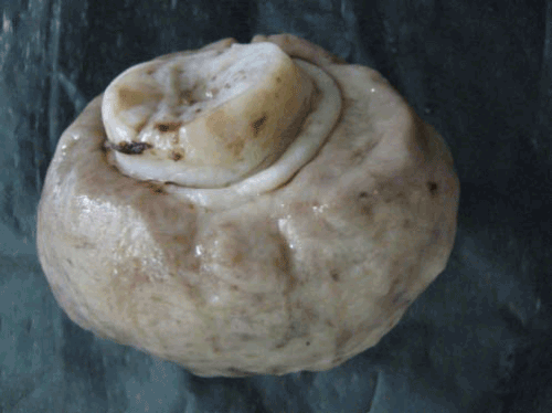
| Received single, soft polypoid, pedunculated tumor mass. External surface was smooth and glistening. Tumor mass was measuring 6.5 x 5.5 x 4 cm (Figure 1). Cut surface was gelatinous, glistening, slimy, homogenous, grayish white, solid with some cystic areas. |
| |
|
|
Figure 1: Gross specimen showing polypoid, pedunculated, glistening, grayish white tumor mass. |
|
| |
| Microscopy |
| |
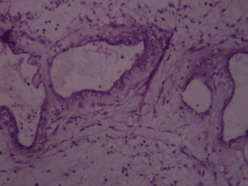
| Sections revealed poorly circumscribed tumor mass, partly covered by ectocervical stratified squamous epithelium. The tumor mass was composed of spindle and stellate shaped cells with ill-defined cytoplasmic borders, numerous, variable sized thick muscular and thin walled blood vessels, embedded in the abundant myxoid stroma (Figure 2). Occasional endocervical gland was also present, in the tumor mass. Cellular atypia was not seen. No cellular pleomorphism, anisonucleosis, increased mitotic activity or necrosis were seen. No lipoblasts or nerve sheath elements were present. Histological diagnosis of aggressive angiomyxoma of cervix was given. |
| |
|
|
Figure 2: Photomicrograph showing blood vessels of variable caliber, in a myxoid stroma (H& E, X 100). |
|
| |
| Discussion |
| |
| Aggressive angiomyxoma is a slowly growing myxoid neoplasm that occurs chiefly in the genital, perineal and pelvic regions of adult women. The neoplasm predominantly affects reproductive age females with peak incidence during third decade of life. The female to male ratio is 6:1. In women vulvar region is the most common site of involvement [3]. In the reviewed literature, a rare case of AAM mimicking Cervical polyp has been reported by Paplomata E et al [4]. Our case may be the second rare case of AAM presenting as a cervical polyp. Our patient was |
| |
| 45- year- old female. Size of the tumor was more than 6 cm in diameter. The tumor presented as slowly growing, painless, polypoid, pedunculated mass. Microscopic examination showed poorly circumscribed tumor mass composed of variable sized thick and thin walled blood vessels, abundant myxoid stroma and uniform bland spindle and stellate cells. Considering all these findings, histopathological diagnosis of aggressive angiomyxoma of cervix was given. Aggressive angiomyxoma must be distinguished from the more common benign and malignant myxoid tumors including myxoma, myxoid liposarcoma, myxoid neurofibroma, myxoid leiomyoma, leiomyosarcoma, myxoid liposarcoma, myxoid malignant fibrous histiocytoma and botryoides rhabdomyosarcoma. AAM may also be clinically misdiagnosed as polyps, myxoma, lipoma and Bartholin’s cyst of vagina. The diagnosis of angiomyxoma may be difficult to establish. The distinctively striking vascular component in AAM helps to rule out the above mentioned neoplasm as differential [5]. In our case, considering the location of lesion, two close differential diagnoses were superficial angiomyxoma and angiomyofibroblastoma of cervix which were ruled out histologically. Superficial angiomyxomas arise most often in the head and neck region and occasionally in the vulvovaginal region. They are slowly growing and circumscribed nodules. They are usually less than 3-4 cm in diameter. Histologically multiloculated, poorly delimited myxoid mass composed of plump spindle cells, numerous thin walled blood vessels and inflammatory cells. Thick walled blood vessels are not seen in superficial angiomyxoma. These tumors have potential for local nondestructive recurrence in approximately 30% cases [6]. Angiomyofibroblastomas are well circumscribed, round, ovoid or lobulated, usually less than 3 cm in diameter. Majority of them measure 2- 8 cm. The cut surface is gray pink to yellowish brown to tan. There are hyper & hypocellular areas. Perivascular hypercellularity is present. Epithelioid plump spindle cells, multinucleate cells and many thin walled blood vessels are present. Recurrence is rare [3,6]. Babala P et al. reported a case of angiomyofibroblastoma of the cervix uteri. Recognition of this entity is important to avoid misdiagnosis of the other angiomyxoid neoplasms [7]. Complete surgical excision is the gold standard for AAM because of its tendency to recur locally. The recurrence rate varies from 36-70%. Surgery causes significant morbidity due to its frequent occurrence in lower pelvis and perineum with proximity to genitourinary and anorectal structures. Most surgeons aim at complete resection (wide excision with tumor free margin), incomplete or partial resection is acceptable when high operative morbidity is anticipated and |
| |
| Fertility is an issue. Treatment options include use of hormonal manipulation such as tamoxifen, raloxifen or GnRH analogs, radiotherapy and arterial embolisation [4,8]. It is typically a benign, non-metastatising neoplasm. In two cases however multiple metastasis have been reported [9,10]. So long term follow-up of patient is necessary. Our patient is followed up for six months after surgery. There is no evidence of recurrence till date. |
| |
| |
| References |
| |
- Steeper TA, Rosai J (1983) Aggressive angiomyxoma of female pelvis and perineum. Report of nine cases of a distinct type of gynecologic soft tissue neoplasm. Am J Surg Pathol 7: 463-475.
- Behrnwala KA, Thomas JM (2003) Aggressive angiomyxoma: A distinct clinical entity. Eur J Surg Oncol 29: 559-563.
- Abdul-Karim KW, Chang AE, Fletcher JA, Folpe AL, Geisinger KR, et al. (2001) Benign soft tissue tumors and pseudotumors of miscellaneous type. Weiss SW, Goldblum JR. (4th edn) Enzinger Weiss’s Soft tissue tumors. Mosby St Louis, p: 1419-1481.
- Paplomata E, Fotas A, Balaxis D, Filindris T, Charalambous S, Rombis V (2010) Aggressive angiomyxoma mimicking cervical polyp. Pelviperineology 29: 30-31.
- Akbulut M, Demircan NC, Colakoglu N, Duzcan E (2006) Aggressive angiomyxoma of the vulva: A case report and review of literature. Aegean Pathology Journal 3: 1-4.
- Fletcher M, Christopher D (2007) Soft tissue tumors. (3rd edition), Diagnostic Histopathology of tumors. Churchill Livingstone, Elsevier: 1527- 1592.
- Babala P, Biro C, Klacko M, Miklos P, Ondrus D (2011) Angiomyxofibroblastoma of the Cervix Uteri: A Case Report. Klin Onkol 24: 133-136.
- Rudra S, Banerji RN, Mani NS (2007) Case Report - Aggressive Angiomyxoma. MJAFI 63: 386-387.
- Siassi RM, Papadopoulos T, Matzel KE (1999) Metastasizing aggressive angiomyxoma. N Eng J Med 341: 1772.
- Blandamura S, Cruz J, Faure Vergara L, Machad Puerto I, Ninfo V (2003) Aggressive angiomyxoma: a second case of metastasis with patient’s death. Human Pathol 34: 1072-1074.
|
| |
| |