|
|
| Abdul Halim Shibghatullah1, Mohamad Khir Abdullah1 and Irfan Mohamad2* |
| 1Department of Otorhinolaryngology, Hospital Pakar Sultanah Fatimah, Jalan Salleh, Muar Johor, Malaysia |
| 2Deparment of Otorhinolaryngology-Head & Neck Surgery, School of Medical Sciences, Universiti Sains Malaysia, Kota Bharu, Kelantan, Malaysia |
| *Corresponding authors: |
Dr Irfan Mohamad, MD
M.Med (ORL-HNS)
Dept of Otorhinolaryngology-Head & Neck Surgery
School of Medical Sciences
Universiti Sains Malaysia Health Campus
16150 Kota Bharu, Kelantan, Malaysia
Tel: 609-7664156
E-mail: irfan@kb.usm.my |
|
| |
| Received April 02, 2012; Published August 26, 2012 |
| |
| Citation: Shibghatullah AH, Abdullah MK, Mohamad I (2012) Unusual Superficial Lateral Course of Sigmoid Sinus. 1: 250. doi:10.4172/scientificreports.250 |
| |
| Copyright: © 2012 Shibghatullah AH, et al. This is an open-access article distributed under the terms of the Creative Commons Attribution License, which permits unrestricted use, distribution, and reproduction in any medium, provided the original author and source are credited. |
| |
| Abstract |
| |
| Sigmoid sinus is an important landmark in mastoidectomy surgery. It serves the posterior border of mastoid antrum and drilling was done anterior to it. Superiorly, it meets the dura forming the sinodural angle. We report a case of a gentleman who was presented with otalgia and headache associated with altered conscious level. As the medical management failed to show improvement, exploratory mastoidectomy was done. We found the unusual location of sigmoid sinus which was located more anterior and superficial than normal site. |
| |
| Keywords |
| |
| Mastoidectomy; Sigmoid sinus; Superficial |
| |
| Introduction |
| |
| The variability of sigmoid sinus anatomy was well described in the literature. In addition, some of them may be very superficial as in our case. Thus, its relationship with other surgical landmarks is also variable. Surgeons should be well versed of these varied positions in order to avoid unwanted effect during mastoidectomy surgery. |
| |
| Case Summary |
| |
| A 46-year-old gentleman was referred to otorhinolaryngologist with history of right earache for 1 month duration. He was prescribed with oral antibiotic by his family doctor but he did not complete the course. The problem was getting worse for the last four days. The earache was associated with occipital headache, fever, vomiting and deteriorating level of consciousness. On examination, he was restless with Glasgow Coma Scale of 10/15, tachycardia and having low grade temperature. |
| |
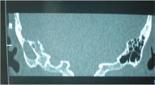
| Otoscopic examination showed retracted right tympanic membrane without any discharge seen. Other ENT and systemic examinations were unremarkable. Computed tomographic scan showed sclerosis of right mastoid bone. The remaining of mastoid air cells were filled with fluid (Figure 1). The right middle ear cavity also filled with fluid. The roof of the middle ear attic which also forms the floor of the right middle cranial fossa was breached, creating a communication between the middle ear and the intracranial cavity. However, no intracranial focal enhancing lesion was seen. |
| |
|
|
Figure 1: Coronal CT scan showed fluid density filled the air cells. |
|
| |
| He was intubated and started with intravenous Rocephine, Gentamycin, Cloxacillin and Metronidazole. On day 7, as patient condition didn’t show any significant improvement, exploratory mastoidectomy was planned. |
| |
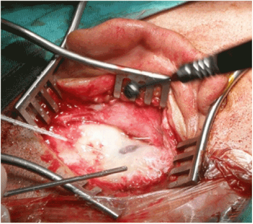
| Intraoperatively, a bluish discoloration of the cortex was identified after raising the post auricular flap (Figure 2). After minimal drilling of the cortex, sigmoid sinus emerged. It was very superficially located. It was identified anterior to the mastoid antrum. The number of air cells was small. There was minimal sign of inflammation. No focal infection was identified. Myringotomy was performed but dry in tapping. |
| |
|
|
Figure 2: Sigmoid sinus was identified after minimal drilling of cortex. |
|
| |
| Post operatively patient showed significant improvement. The patient regained consciousness. The fever resolved. |
| |
| Discussion |
| |
| In a simple or cortical mastoidectomy, the aim of the procedure is to convert the mastoid air cells into a single cavity. One of the indications is acute mastoiditis which is not resolved with medical management. Although most cases of acute mastoiditis can now be managed medically with the development of newer antibiotics, morbidity, mortality and complications of this disease continue to be reported. In our case, the patient was clinically and radiologically diagnosed as acute mastoiditis with intracranial complication. Thus, starting him on antibiotics was the first option. Nevertheless, the patient showed no improvement despite ample period was given for the drugs to take its effect. |
| |
| Post auricular incision provides wide field exposure for visualization of important anatomical landmarks. After removal of cortex, in the normal mastoid, the sigmoid sinus can be identified by its prominent bony bulge into the mastoid and by its bluish colour [1]. Despite of this, the wide variety of sigmoid sinus location in the temporal bone has been reported [2-4]. |
| |
| The relationship between anatomical variations of sigmoid sinus and the degree of mastoid pneumatization has been established. By using high resolution CT scan, Ichijo et al found that the distance between the posterior wall of the external auditory canal and the anterior edge of the sigmoid sinus was shorter in poorly a pneumatized mastoid [5]. Similarly Shatz and Sade (1990) found a significant relationship between the degree of mastoid pneumatization and the distance of the sigmoid sinus from the posterior border of the external ear canal in the pneumatized mastoids [6]. They also found that a significance difference between non-pneumatized and pneumatized mastoids regarding the distance of sigmoid sinus from the posterior border of the external ear canal. A Aslan et al (1996) studied regional mastoid pneumatization in 25 temporal bones found that pneumatization in the area of sinodural angle were significantly related with the position of sigmoid sinus [7]. |
| |
| By using seven reference points, surgical classifications of the location of sigmoid sinus were proposed. Based on the 96 temporal bone dissection performed, they were grouped into 3 types. In type 1, the location of the sigmoid sinus was posterior, enlarging the Trautmann’s triangle. In type 2 (the most common), the sigmoid sinus was located anteriorly diminishing the size of Trautmann’s triangle. In the type 3, the sigmoid sinus was medially displaced which also reduced the area of Trautmann’s triangle [8]. In our case, the sigmoid sinus most probably was the type 2 in anteroposterior axis but very superficial (lateral) in term of depth. |
| |
| |
| References |
| |
- Nadol JB, Schuknecht HF (1993) Surgery of the Ear and Temporal bone, Raven Press Ltd, 145-154.
- Glasscock ME, Shambaugh GE (1990) Surgery of the Ear. Philadelphia: Sauders.
- Sanna M (1995) Atlas of temporal Bone and Lateral Skull Base Surgery. Thieme.
- Tos M (1995) Manual of Middle Ear Surgery: Mastoid and Reconstructive Procedures. Thieme Vol.3.
- Ichijo H, Hosokawa M, Shinkawa H (1996) The relationship between mastoid pneumatization and the position of the sigmoid sinus. Eur Arch Otorhinolaryngol 253: 421-4.
- Shatz A, Sade J (1990) Correlation between mastoid pneumatization and position of the lateral sinus. Ann Otol Rhinol Laryngol 99: 142-145.
- Aslan A, Kobayashi T, Diop D, Balyan FR, Russo A, et al. (1996) Anatomical Relantionship Between Position of the Sigmoid Sinus and Regional Mastoid Pneumatization. Eur Arch Otorhinolaryngol 253: 450-453.
- Sarmiento PB, Eslait FG (2004) Surgical classification of variations in the anatomy of sigmoid sinus. Otolaryngol Head Neck Surg 131: 192-9.
|
| |
| |


