| Research Article |
Open Access |
|
| Shane A O’Hanlon1*, Valerie Lim2, Norman Delanty3 and Richard Liston4 |
| 1Kerry General Hospital, Kerry, Ireland, Spr in Geriatric Medicine |
| 2The Prince Charles Hospital, Brisbane, Queensland, Australia, Medical Registrar |
| 3Beaumont Hospital, Dublin, Ireland, Consultant Neurologist |
| 4Kerry General Hospital, Kerry, Ireland, Consultant Geriatrician |
| *Corresponding authors: |
Shane O’Hanlon
Graduate Entry Medical School
University of Limerick, Ireland
Tel: +353 85 1414460
E-mail: sohanlon@gmail.com |
|
| |
| Received November 15, 2011; Published August 29, 2012 |
| |
| Citation: O’Hanlon SA, Lim V, Delanty N, Liston R (2012) Status Epilepticus in Older Patients. 1: 260. doi:10.4172/scientificreports.260 |
| |
| Copyright: © 2012 O’Hanlon SA, et al. This is an open-access article distributed under the terms of the Creative Commons Attribution License, which permits unrestricted use, distribution, and reproduction in any medium, provided the original author and source are credited. |
| |
| Abstract |
| |
| Status Epilepticus (SE) is an important medical emergency. There are several challenges when treating older people, including increased mortality, lack of evidence for treatment options, and a high rate of adverse effects. There are few studies focusing on management of SE in older people. This review highlights aspects of care in older patients, provides treatment guidelines and points out deficiencies in the evidence base. There is a need for more clinical research in this area. |
| |
| Introduction |
| |
| Status Epilepticus (SE) is a medical emergency involving persistent seizure activity. In adults it is more common in older age and has highest mortality in this group [1-6]. It requires urgent and aggressive treatment to prevent serious sequelae including disability and death. The prolonged post-ictal state seen in old age can add further hazards. SE occurs in patients with pre-existing epilepsy but also presents as a result of other illness. Tallis notes that seizures in older people have a significant psychological effect; by undermining self-confidence at the deepest level, it may seem a “harbinger of death.” |
| |
| Methods |
| |
| A literature review was carried out to identify trials that studied status epilepticus in older patients. The Medline database was searched using MeSH terms “Status epilepticus”, “Aged” and “Aged, 80 and over”, and also using the terms “elderly” and “older”. Searches were also performed on the EMBASE and Web of Science databases, the Cochrane collaboration database and using the Google search engine [7-10]. Hand searches were completed on relevant journals in the areas of Neurology and Geriatric Medicine. References in all articles were checked to find other possible studies to include. All relevant results were reviewed independently by two authors and information drawn from all suitable articles. |
| |
| Definition |
| |
| Status epilepticus was originally defined by Gastaut as a seizure persisting “for a sufficient length of time or repeated frequently enough to produce a fixed or enduring epileptic condition”. In later definitions a timeframe was applied: “a continuous seizure lasting more than 30 minutes, or two or more seizures without full recovery of consciousness between any of them”. This was thought to reflect time to pathophysiological damage. However in clinical practice a seizure lasting longer than 5 minutes should be assumed to be status epilepticus [11-16]. This is because the average duration of a generalised tonic-clonic seizure is usually around 60 seconds and is almost always less than 2 minutes. If it persists longer then it is unlikely to resolve spontaneously and is probably status epilepticus. It should thus be treated as such. |
| |
| Clinical features |
| |
| SE was classified by the International League Against Epilepsy into partial and generalised. A simpler clinical classification is either convulsive or non-convulsive [17]. The patient with non-convulsive status epilepticus may present with any of a wide range or symptoms which are discussed below. |
| |
| • Generalised Convulsive Status Epilepticus (GCSE) |
| |
| This is the most common form, and there is little difficulty making the diagnosis clinically. The patient has tonic-clonic convulsions and usually a tachycardia, high temperature, unstable blood pressure and may have incontinence, vomiting and cardiac arrhythmias [18]. It is worth being aware of the other subtypes, which are less easily recognised. |
| |
| • Non-Convulsive Status Epilepticus (NCSE) |
| |
| NCSE consists of a disparate group of entities, which vary widely in aetiology and prognosis. They tend to lack the typical features of seizure activity and may present clinically as confusion, altered behaviour or even coma (See Box 1). They may evolve from convulsive SE or arise de novo [19-25]. In a study of generalised electrographic SE, 40% of patients continued to have subclinical SE, despite the clinical impression that seizures had stopped. EEG confirmation is essential. NCSE is classified into complex or simple partial status epilepticus (CPSE and SPSE) as well as absence status (AS). In older people it most commonly presents as CPSE. CPSE tends to recur and may be difficult to treat, with a response to intravenous benzodiazepines in only about 60% of patients. Cognitive and behavioral sequelae have also been reported, underlining the need for early recognition and treatment. Mortality may reach up to 26.3% in older people [26-30]. |
| |
|
|
Box 1: Features that may indicate NCSE |
|
| |
| NCSE accounts for about 4-20% of all cases of SE. One case control study in older patients noted that only one third of the patients with NCSE had a history of epilepsy. Compared to controls that had altered mental status but no EEG evidence of NCSE, they were more likely to have a history of dementia (in almost two thirds) or stroke (in one third). Those who had NCSE also had a longer mean hospitalisation (25 days vs 7 days) and were more likely to have a worse outcome (50% vs 5.8%). Patients were mostly female as in most studies [31]. |
| |
| NCSE in critically ill elderly patients has been shown to have high mortality of about 50%. Veterans Affairs studies found that 65% of the patients with NCSE died within 30 days of an episode compared to 27% of patients with GCSE. Another retrospective study of 100 patients found that mortality rates were higher in patients with acute medical conditions (27%) compared to patients with history of epilepsy (3%) and patients with cryptogenic (18%) causes of NCSE; and in patients with severe mental status impairment compared to those with mild impairment. Those who developed complications of treatment were also more likely to die [32]. The authors recommended that patients with epilepsy as the only cause of NCSE should not be routinely treated very aggressively because patients were more likely to die from the complications of treatment than the NCSE itself; and that in patients with acute medical aetiologies and cryptogenic causes of NCSE, aggressive treatment is warranted. |
| |
| • Absence SE |
| |
| Absence status is a prolonged, generalized, and nonconvulsive seizure characterized by impairment of consciousness at times associated with other clinical manifestations such as automatisms or subtle myoclonic, tonic, atonic, or autonomic phenomena [33]. In older patients it has been reported to occur as situation-related phenomenon. It may occur in any epilepsy that involves typical absences, but may also present de novo. AS can occur spontaneously or as a consequence of drug withdrawal, or other external factors such as sleep deprivation, excessive alcohol intake, or metabolic disturbances. The prognosis of “de novo” absence status is generally good and recurrences are rare. |
| |
| • Epilepsia Partialis Continua |
| |
| Also referred to as partial motor status, epilepsia partialis continua (EPC) is classified under the group of focal SE. It was originally described in the 19th century by Koshewnikow. The clinical presentation is of repetitive myoclonic jerks with hemiparesis [34-39]. There is more frequent involvement of the upper half of the body. The origin of the seizures is thought to be the primary motor area. The commonest cause appears to be vascular disorders though encephalitis and neoplasms should be considered. There is little information on EPC in older patients, though it has been reported to be mistaken for Parkinson’s disease and has been described as a result of vasospasm from subarachnoid haemorrhage in a 72 year old, responsive to valproate. |
| |
| • Psychogenic SE |
| |
| Psychogenic non-epileptic seizures (PNES) are characterized by episodes of altered behaviour which may appear similar to epileptic seizures (see Box 2 for how to differentiate). PNES is classified by DSMIV as a conversion disorder; symptoms have no organic cause but are not intentionally produced. They do not usually occur for the first time in old age. Up to 30% of patients with PNES also have epilepsy [40-43]. An important differential diagnosis is frontal lobe status epilepticus. Video EEG monitoring is the gold standard in establishing the diagnosis. Delay in diagnosis occurs in almost all cases, and leads to iatrogenic complications due to inappropriate AED treatment. PNES status epilepticus has been reported to occur in up to 77% of PNES patients, though these patients tended to be of a young age. |
| |
|
|
Box 2: Features suggestive of Psychogenic Seizures |
|
| |
| Epidemiology |
| |
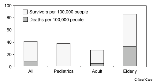
| There is a continuing rise in the annual incidence of epilepsy in old age. For status epilepticus, there is a bimodal peak in incidence – in young children and in people aged 60 and over [44]. The incidence of SE in the elderly is almost twice that of the general population at 86 per 100,000 per year (Figure 1). This may be an underestimate as patients may die out of hospital due to SE, or be misdiagnosed as other conditions before death in hospital. As the population ages in Western countries, it is expected that its incidence will rise. Up to 30% of adult patients with a new diagnosis of epilepsy first present in SE, and in older patients, every third acute symptomatic seizure is status epilepticus (Figure 1). |
| |
|
|
Figure 1: A graphical representation of mortality and incidence for four population groups. Figure reproduced with permission from Bassin, 2002. |
|
| |
| Aetiology |
| |
| Over half of patients with SE do not have a diagnosis of epilepsy and often it is precipitated by an acute illness. In one study of hospitalised patients with a median age of 65 years, de novo status epilepticus was most commonly caused by stroke and metabolic derangements [45- 49]. De Lorenzo’s review of common causes of SE in older patients also emphasises the importance of cerebrovascular disease as a factor in SE (Table 1 and 2). Alcohol intoxication or withdrawal is less commonly a cause in older patients but should still be considered. In some cases no obvious cause may be found. There is also an association with advanced Alzheimer's disease, which has been identified as a risk factor for newonset generalized tonic-clonic seizures in older adults. |
| |
|
|
Table 1: Causes of epilepsy in older patients |
|
| |
|
|
Table 2: Causes of SE in older patients |
|
| |
| Investigation |
| |
| Blood investigations should include full blood count and CRP to check for infection, biochemistry for electrolyte abnormalities, calcium and glucose level [50]. CK rises with seizure activity. Prolactin is not useful in status epilepticus as it returns to normal with persistent seizures. An arterial blood gas will reveal hypoxia or evidence of metabolic disarray. Antiepileptic drug levels should be assayed if the patient has been prescribed them, and liver function checked. A toxicology screen may be useful [51]. |
| |
| Brain imaging is mandatory, with either CT or MRI. ECG helps to rule out cardiac arrhythmias such as long QT syndrome which may present as seizures and a lumbar puncture is indicated if central nervous system infection is possible. In the event of doubt about the diagnosis an EEG may help to clarify the matter. It is also useful for diagnosis of non-convulsive SE, which may develop from convulsive SE but be easily missed by the clinician. One EEG study found that normal variant patterns considered as epileptiform abnormalities, or non-specific abnormalities are more frequently recorded in older age [52-54]. However a later study comparing changes on EEGs in young patients (20 to 59 years) and older patients (over 60 years of age) who had epilepsy found decreased background rhythm, rhythmicity, and amplitude in the older group. |
| |
| Initial treatment |
| |
| Early treatment is important as longer seizure duration leads to less response to treatment and more neuronal damage. The priorities are to stop the seizure and apply basic life support immediately. Airway, breathing and circulation should be assessed and intervention made if necessary. The patient should be kept in a safe area away from objects that may cause injury if struck [55]. Nothing should be put in the mouth. IV access should be established. A fingerprick test for blood glucose level should be performed, and IV dextrose given if necessary. Thiamine should be administered where malnutrition is suspected. |
| |
| Pharmacologic therapy |
| |
| Immediate treatment is initially with a benzodiazepine. Lorazepam is the preferred first line agent due to its favourable pharmacokinetics. It rapidly achieves high concentration in the brain and appears to have a lower risk of seizure recurrence than diazepam [56-58]. A Cochrane review found that lorazepam is better than diazepam or phenytoin alone for cessation of seizures and carries a lower risk of continuation of status epilepticus requiring a different drug or general anaesthesia. The incidence of respiratory depression is higher in older people. One small prospective observational study suggested that in critically ill older patients with NCSE, giving intravenous benzodiazepines was associated with increased risk of death. |
| |
| Second line therapy |
| |
| Most guidelines recommend intravenous phenytoin as second line if benzodiazepines have failed. It has established efficacy and its side effects are known. It may produce hypotension and cardiac arrhythmias and cardiac monitoring is recommended. These problems are seen more often in patients over 50 years [59-63]. The infusion site reaction called “purple glove syndrome” is thought to be due to local thrombosis. It is less common if using the prodrug fosphenytoin, but this is more expensive. Fosphenytoin can be given intramuscularly if intravenous access is difficult to establish. Sodium valproate has been suggested as an alternative second line agent, but is approved for this in some countries only. Two small randomised comparative studies of phenytoin and valproate favoured valproate. , Valproate also appears to have a low incidence of cardiovascular side effects [64]. |
| |
| In older people, there appears to be no uncontroversial drug of first choice. Age-related changes such as progressive decline in AED protein binding by albumin, increased volume of distribution, and slow elimination can affect pharmacokinetics and pharmacodynamics. Older people are often on other medications, which makes drug interaction more likely. They appear to be more responsive to antiepileptic drug therapy than younger groups, but are also more likely to experience side effects at lower serum antiepileptic drug concentrations. A 1994 trial found a mean side effect rate of 20% with valproate or phenytoin [65-67]. Just over 20% of patients were not satisfactorily controlled. The choice of an AED is also made difficult by the paucity of evidence based data on their use in elderly patients, especially in the very old and those with comorbidities. Recent small case series have suggested that intravenous levetiracetam may be useful in older patients. No trials were found comparing treatment options for SE in younger and older patients. Trials comparing different treatments tended to have a younger cohort than would be generally seen in clinical practice [68]. |
| |
| As is standard practice in Geriatric Medicine, the treatment should be tailored to the patient. For example, antacids and enteral feeding decrease the absorption and efficacy of phenytoin. Valproate levels may be decreased and phenytoin levels may be increased in patients taking both medications. Carbamazepine is metabolized in the liver, and as hepatic function can be reduced in older people, levels should be monitored closely. It also has a greater association with hyponatraemia in old age. The VA Cooperative Study on epilepsy in the elderly demonstrated that lamotrigine and gabapentin were better tolerated than carbamazepine [69-71]. Newer drugs include topiramate and lacosamide but experience is limited. Topiramate has been reported to have adverse effects on cognition, even in younger patients, which leads to a high discontinuation rate. |
| |
| Refractory SE |
| |
| In some cases SE does not resolve despite the application of first and second line treatments. It is then termed refractory SE. Anaesthetic agents (such as pentobarbital, propofol, or midazolam) and mechanical ventilation may be necessary. It is estimated that SE becomes refractory in 9% to 40% of cases. Mortality for RSE is as high as 48% and only about a third of patients return to their premorbid functional baseline [72]. |
| |
| ICU management |
| |
| Ala-Kokko looked at incidence of infections in patients with status epilepticus requiring intensive care and effect on resource utilization. He concluded that the infection rate of status epilepticus patients was high and nosocomial infections were associated with more severe illness, treatment escalation, prolonged hospital stay and enhanced resource utilization. In one study of critically ill older patients with NCSE, those who were managed outside of ICU had significantly shorter hospital stays (22.3 vs. 39.2 days) [73-76]. |
| |
| Surgical management |
| |
| A small range of case reports and series has reported the use of surgery such as temporal lobectomy in the management of epilepsy, however this is infrequent in status epilepticus and the oldest patient reported was 45 years [77-79]. |
| |
| Out of hospital management |
| |
| Older patients who need to be treated in the community or in long stay facilities where intravenous drugs may not be available will nevertheless require immediate treatment. Rectal diazepam has been successfully used and is an easy and well tolerated treatment. In one study with a mean patient age of around 50 years, out of hospital administration of intravenous lorazepam, diazepam or placebo were compared. The treatment group showed a clear benefit, with lorazepam slightly better. The rate of respiratory complications was also less in the treatment group, which may suggest that SE itself causes more respiratory effects than the treatment. However 41 to 57% of patients were still in SE at the time of arrival to the Emergency Department. Lorazepam can also be given intramuscularly but it needs to be kept refrigerated, limiting its availability. Another study demonstrated that the administration of a single dose of diazepam rectal gel was safe and effective in the treatment of homebound patients who had repetitive seizures. Newer formulations such as buccal midazolam have been proposed but no randomised controlled trials have been published in older adults. Comparisons with diazepam in children have had positive results, and it may be useful in situations where IV access is difficult [80]. |
| |
| Maintenance therapy |
| |
| Once instituted, AED therapy in older people is often lifelong. Due to the permanent nature of the commonest causes of epilepsy in older people, withdrawal is not usually recommended. In acute symptomatic SE where the provoking circumstance is corrected (e.g. hyponatraemia), AEDs can be slowly withdrawn once the patient remains well. The dosages recommended for the adult population may not be suitable for the elderly. A standard “dose for elderly” is not likely to be useful in all patients, and there can be large variance in therapeutic doses among older people. A general strategy is start low and go slow, considering the individual. Some studies suggested increased cognitive dysfunction in older patients, but Craig and Tallis found that there were no significant differences in either the general adult population or older people [81]. |
| |
| Driving |
| |
| The regulations governing driving for those diagnosed with epilepsy vary by country. In the UK, the Driver and Vehicle Licencing Authority must be notified, and driving is restricted until the authority allows [82]. The responsibility lies with the licence-holder to inform the DVLA, but doctors also have a public duty of care to inform the authority. For newly diagnosed epilepsy, the regulations provide for a driving ban until one year after last seizure. |
| |
| Morbidity and mortality |
| |
| The overall mortality rate for SE is 20% however it rises with age and can be as high as 80% in older groups. In a 5 year review of almost 12,000 cases of SE in the US, the mortality in those over 80 years was over eight times that of patients aged in their twenties. Mortality rose progressively in each decade from age 21. The higher mortality in older patients may be due to a higher incidence of stroke and tumors in this age group, which have a poorer prognosis than causes which occur commonly in younger people. Knake found that SE after stroke is associated with increased long-term case fatality and that the occurrence of SE in patients with cerebrovascular disease indicates a high risk of death within 3 years [83]. It has also been suggested that these patients may be more inclined to develop complications such as pneumonia or venous thrombosis (see Box 3), and that the comorbidities are higher in older people. There has been some debate as to whether status epilepticus itself causes cognitive decline. It has not been ascertained whether the seizures are caused by the cognitive impairment or vice versa. |
| |
|
|
Box 3: Complications of SE |
|
| |
| Prognosis |
| |
| The prognosis is related to the aetiology and duration of the seizures. For example, when SE complicates acute ischaemic stroke, mortality is 3 times higher than in stroke alone [17]. Primary predictors of poor outcome in SE are anoxia, prolonged seizure (greater than 1 hour), advanced age and severe physiological disturbances. Kaplan reviewed the prognosis of NCSE and suggested that the it depends not only on NCSE type but also on level of consciousness [83]. |
| |
| One should also bear in mind that even the diagnosis of epilepsy in older people can be severely limiting on lifestyle, with reduced confidence, mobility and independence. This can trigger further decline and deconditioning, and a multidisciplinary assessment should be undertaken. For example, Tallis recommends an occupational therapist assessment of the home for potential sources of danger, and a physiotherapist assessment to encourage maintenance of mobility. An epilepsy nurse specialist may be available in some centres to provide support and advice. Factors which precipitate fits, such as lack of sleep or drinking alcohol should be avoided. Patients should be monitored for bone loss which can occur due to some AEDs. There is a high risk of falls in patients with epilepsy. Concordance should be achieved between patient and clinician to ensure a satisfactory overall management plan is instituted. |
| |
| Recurrence |
| |
| Recurrence of SE occurs in 13.3–18.5% of cases overall. It depends on the underlying cause for the seizures. In adults, almost all (93.8%) patients with recurrences have a history of epilepsy.81 Old age was found to be a significant predictor of seizure recurrence by the FIRST seizure trial group.82 |
| |
| Conclusion |
| |
| Status epilepticus is a significant cause of morbidity and mortality in older patients. It is underrecognised, particularly the non-convulsive form. Older age affects incidence, aetiology and mortality of status epilepticus. Cerebrovascular disease is a common association in older people. There is a lack of study of treatment options in this group. The optimal management for older people has not yet been elucidated. A higher incidence of medication side effects is also a concern. Due to lack of evidence, and unpredictable pharmacologic effects, it is important to tailor therapy to each patient. Status epilepticus in older people needs closer examination and further clinical research. The distinctive aetiologies and presentation of SE in older patients adds to the difficulty in appropriate diagnosis and management. |
| |
| |
| References |
| |
- Chin RF, Neville BG, Scott RC (2004) A systematic review of the epidemiology of status epilepticus. Eur J Neurol 11: 800-810
- Godfrey JW, Roberts MA, Caird FI (1982) Epileptic seizures in the elderly: II. Diagnostic problems. Age Ageing 11: 29-34.
- Shorvon S (2001) The management of status epilepticus. J Neurol Neurosurg Psychiatry. 2: 1122-1127.
- Grimley Evans J, Williams TF, et al. (2000) editors. Oxford textbook of Geriatric Medicine. 2nd ed. USA: Oxford University Press 769.
- Gastaut H (1973) Dictionary of epilepsy. Part 1. Definitions. World Health Organization, Geneva 72.
- (1993) Treatment of convulsive status epilepticus. Recommendations of the Epilepsy Foundation of America’s Working Group on Status Epilepticus. JAMA 270: 854–859.
- Lowenstein DH, Bleck T, Macdonald RL (1999) It's time to revise the definition of status epilepticus. Epilepsia 40: 120-122.
- Theodore WH, Porter RJ, Penry JK (1983) Complex partial seizures: clinical characteristics and differential diagnosis. Neurology 33: 1115–1121
- Drislane FW, Schomer DL (1994) Clinical implications of generalized electrographic status epilepticus. Epilepsy Res 19: 111-121.
- Sheth RD, Drazkowski JF, Sirven JI, Gidal BE, Hermann BP (2006) Protracted ictal confusion in elderly patients. Arch Neurol 63: 529-532.
- Tomson T, Lindblom U, Nilson BY (1992) Nonconvulsive status epilepticus in adults: thirty-two consecutive patients from a general hospital population. Epilepsia 33: 829-835.
- Cockerell OC, Walker MC, Sander JW, Shorvon SD (1994) Complex partial status epilepticus: a recurrent problem. J Neurol Neurosurg Psychiatry 57: 835-837.
- Ferlazzo E (2010) Confusional status epilepticus in elderly. BMC Geriatrics 10: L35.
- Walker M, Cross H, Smith S, Young C, Aicardi J, et al. (2005) Nonconvulsive status epilepticus: Epilepsy Research Foundation workshop reports. Epileptic Disord 7: 253-296.
- Bottaro FJ, Martinez OA, Pardal MM, Bruetman JE, Reisin RC (2007) Nonconvulsive status epilepticus in the elderly: a case-control study. Epilepsi 48: 966-972.
- Kaplan PW (2005) The clinical features, diagnosis, and prognosis of nonconvulsive status epilepticus. Neurologist 11: 348-361.
- Husain AM, Horn GJ, Jacobson MP (2003) Non-convulsive status epilepticus: usefulness of clinical features in selecting patients for urgent EEG. J Neurol Neurosurg Psychiatry 74: 189-191
- Litt B, Wityk RJ, Hertz SH, Mullen PD, Weiss H, et al. (1998) Nonconvulsive status epilepticus in the critically ill elderly. Epilepsia 39: 1194-1202.
- Treiman DM, Meyers PD, Walton NY, Collins JF, Colling C, et al. (1998) A comparison of four treatments for generalized convulsive status epilepticus. Veterans Affairs Status Epilepticus Cooperative Study Group. N Engl J Med 339: 792-798.
- Shneker BF, Fountain NB (2003) Assessment of acute morbidity and mortality in nonconvulsive status epilepticus. Neurology 61: 1066-1073.
- Thomas P, Valton P, Genton P (2006) Absence and myoclonic status epilepticus precipitated by antiepileptic drugs in idiopathic generalized epilepsy. Brain 129: 1281-1292.
- Thomas P, Beaumanoir A, Genton P, Dolisi C, Chatel M (1992) “De novo” absence status of late onset: report of 11 cases. Neurology 42: 104-110.
- Thomas P, Andermann F () Late-onset absence status is most often situation-related. In Malafosse A, Genton P, Hirsch E, Marescaux C, Broglin D, Bernasconi R (Eds) Idiopathic generalized epilepsies, John Libbey, London 95-09.
- Engel J Jr; International League Against Epilepsy (ILAE) (2001) A proposed diagnostic scheme for people with epileptic seizures and with epilepsy: report of the ILAE Task Force on Classification and Terminology. Epilepsia 42: 796–803.
- Koshewnikow AJ (1895) Eine besondere Form von corticaler Epilepsie. Neurol Centralbl 14: 47-48.
- Bien CG, Elger CE (2008) Epilepsia partialis continua: semiology and differential diagnoses. Epileptic Disord 10: 3-7.
- Sinha S, Satishchandra P (2007) Epilepsia Partialis Continua over last 14 years: experience from a tertiary care center from south India. Epilepsy Res 74: 55-59.
- Al-Hayk K, LeDoux MS (2003) Epilepsia partialis continua mistaken for Parkinson's disease. Mov Disord. 18: 107.
- Rejdak K, Papuc E, Dropko P, Stelmasiak Z (2008) Acute stroke-elicited epilepsia partialis continua responsive to intravenous sodium valproate. Neurol Neurochir Pol 42: 157-160.
- Appleton R, Baker G, Chadwick D (1993) Epilepsy. 3rd ed. Martin Dunitz, London.
- Cragar DE, Berry DT, Fakhoury TA, Cibula JE, Schmitt FA (2002) A review of diagnostic techniques in the differential diagnosis of epileptic and nonepileptic seizures. Neuropsychol Rev 12: 31–64.
- Nandhagopal R (2006) Generalised convulsive status epilepticus: an overview. Postgrad Med J 82: 723-732.
- Reuber M (2008) Psychogenic nonepileptic seizures: answers and questions. Epilepsy Behav 12: 622-635.
- Hovorka J, Nezádal T, Herman E, Nemcová I, Bajacek M (2007) Psychogenic non-epileptic seizures, prospective clinical experience: diagnosis, clinical features, risk factors, psychiatric comorbidity, treatment outcome. Epileptic Disord 9: S52-58.
- Tallis R, Hall G, Craig I, Dean A (1991) How common are epileptic seizures in old age? Age Ageing 20: 442-448.
- DeLorenzo RJ, Hauser WA, Towne AR, Boggs JG, Pellock JM, et al. (1996) A prospective, population-based epidemiologic study of status epilepticus in Richmond, Virginia. Neurology 46: 1029-1035.
- Waterhouse EJ, DeLorenzo RJ (2001) Status epilepticus in older patients: epidemiology and treatment options. Drugs Aging 18: 133-142.
- Sung CY, Chu NS (1989) Status epilepticus in the elderly: etiology, seizure type and outcome. Acta Neurol Scand 80: 51-56.
- Hauser WA (1990) Status epilepticus: epidemiologic considerations. Neurology 2: 9-13.
- Krämer G (2001) Epilepsy in the elderly: some clinical and pharmacotherapeutic aspects. Epilepsia 3: 55-59.
- Bassin S, Smith TL, Bleck TP (2002) Clinical review: status epilepticus. Crit Care 6: 137-142.
- Walker M (2005) Status epilepticus: an evidence based guide. BMJ 331: 673-677.
- Delanty N, French JA, Labar DR, Pedley TA, Rowan AJ (2001) Status epilepticus arising de novo in hospitalized patients: an analysis of 41 patients. Seizure 10: 116–119.
- Romanelli MF, Morris JC, Ashkin K, Coben LA (1990) Advanced Alzheimer's disease is a risk factor for late-onset seizures. Arch Neurol 47: 847-850.
- Tomson T, Lindbom U, Nilsson BY, Svanborg E, Andersson DE (1989) Serum prolactin during status epilepticus. J Neurol Neurosurg Psychiatry 52: 1435-1437.
- Drury I, Beydoun A (1993) Pitfalls of EEG interpretation in epilepsy. Neurol Clin 11: 857-881.
- Hughes JR, Zialcita ML (1999) EEG and epilepsy in the elderly compared to a younger group. Clin Electroencephalogr 30: 126-131.
- Prasad K, Al-Roomi K, Krishnan PR, Sequeira R (2005) Anticonvulsant therapy for status epilepticus. Cochrane Database Syst Rev 19: CD003723.
- Agarwal P, Kumar N, Chandra R, Gupta G, Antony AR, et al. (2007) Randomized study of intravenous valproate and phenytoin in status epilepticus. Seizure 16: 527-532.
- Misra UK, Kalita J, Patel R (2006) Sodium valproate vs phenytoin in status epilepticus: a pilot study. Neurology 67: 340-342.
- Ramsay RE, Rowan AJ, Slater JD, Collins J, Nemire R, et al. (1994) Effect of age on epilepsy and its treatment: results from the VA Cooperative Study [Abstract]. In: Annual meeting of the American Epilepsy Society. New Orleans, Louisiana. Epilepsia 35: 1-173.
- Tallis R, Craig I (1994) Multicentre comparative trial of sodium valproate and phenytoin in elderly patients with newly diagnosed epilepsy. Age Ageing 23: 5.
- Fattouch J, Di Bonaventura C, Casciato S, Bonini F, Petrucci S, et al. (2010) Intravenous Levetiracetam as first-line treatment of status epilepticus in the elderly. Acta Neurol Scand 121: 418-421.
- Aiguabella M, Falip M, Villanueva V, de la Peña P, Molins A, et al. (2011) Efficacy of intravenous levetiracetam as an add-on treatment in status epilepticus: a multicentric observational study. Seizure 20: 60-64.
- Vélez L, Selwa LM (2003) Seizure disorders in the elderly. Am Fam Physician 67: 325-332.
- Sommer BR, Fenn HH (2010) Review of topiramate for the treatment of epilepsy in elderly patients. Clinical Interventions in Aging 5: 88-99.
- Garzon E, Fernandes RM, Sakamoto AC (2003) Analysis of clinical characteristics and risk factors for mortality in human status epilepticus. Seizure 12: 337-345.
- Lowenstein DH, Alldredge BK (1993) Status epilepticus at an urban public hospital in the 1980s. Neurology 43: 483-488.
- Bleck TP (1993) Advances in the management of refractory status epilepticus. Crit Care Med 21: 955-957.
- Jagoda A, Riggio S (1993) Refractory status epilepticus in adults. Ann Emerg Med 22: 1337-1348.
- Mayer SA, Claassen J, Lokin J, Mendelsohn F, Dennis LJ, et al.(2002) Refractory status epilepticus: frequency, risk factors, and impact on outcome. Arch Neurol 59: 205-210.
- Kendall JL, Reynolds M, Goldberg R (1997) Intranasal midazolam in patients with status epilepticus. Ann Emerg Med 29: 415-417.
- Ala-Kokko TI, Säynäjäkangas P, Laurila P, Ohtonen P, Laurila JJ, et al. (2006) Incidence of infections in patients with status epilepticus requiring intensive care and effect on resource utilization. Anaesth Intensive Care 34: 639-644.
- Weimer T, Boling W, Pryputniewicz D, Palade A (2008) Temporal lobectomy for refractory status epilepticus in a case of limbic encephalitis. J Neurosurg 109: 742-745.
- Alldredge BK, Gelb AM, Isaacs SM, Corry MD, Allen F, et al. (2001) A comparison of lorazepam, diazepam, and placebo for the treatment of out-of-hospital status epilepticus. N Engl J Med 345: 631-637.
- Cereghino JJ, Mitchell WG, Murphy J, Kriel RL, Rosenfeld WE, et al. (1998) Treating repetitive seizures with a rectal diazepam formulation: a randomized study. The North American Diastat Study Group. Neurology 51: 1274-1282.
- McMullan J, Sasson C, Pancioli A, Silbergleit R (2010) Midazolam versus diazepam for the treatment of status epilepticus in children and young adults: a meta-analysis. Acad Emerg Med 17: 575-582.
- Talukdar B, Chakrabarty B (2008) Efficacy of buccal midazolam compared to intravenous diazepam in controlling convulsions in children: a randomized controlled trial. Brain Dev 31: 744-749.
- Grimley Evans J, Williams TF, et al. (2000) editors. Oxford textbook of Geriatric Medicine. 2nd ed. USA: Oxford University Press.
- Craig I, Tallis R (1994) The impact of sodium valproate and phenytoin on cognitive function in elderly patients. Epilepsia 35: 381-390.
- https://www.direct.gov.uk/en/Motoring/DriverLicensing/MedicalRulesForDrivers/ MedicalA-Z/DG_185434
- Towne AR (2007) Epidemiology and Outcomes of Status Epilepticus in the elderly. Int Rev Neurobiol 81: 111-127.
- Koubeissi M, Alshekhlee A (2007) In-hospital mortality of generalized convulsive status epilepticus. Neurology 69: 886-893.
- Knake S, Rochon J, Fleischer S, Katsarou N, Back T, et al. (2006) Status epilepticus after stroke is associated with increased long-term case fatality. Epilepsia 47: 2020-2026.
- Rossetti AO, Hurwitz S, Logroscino G, Bromfield EB (2006) Prognosis of status epilepticus: role of aetiology, age, and consciousness impairment at presentation. J Neurol Neurosurg Psychiatry 77: 611-615.
- Helmstaedter C (2007) Cognitive outcome of status epilepticus in adults. Epilepsia 48: 85-90.
- Towne AR (2007) Epidemiology and Outcomes of Status Epilepticus in The Elderly. Int Rev Neurobiol 81: 111-127.
- Tsai1 MH, Chuang YC, Chang HW, Chang1 WN, Lai1 SL, et al. (2009) Factors predictive of outcome in patients with de novo status epilepticus. QJM 102: 57-62.
- Kaplan PW (2003) Nonconvulsive status epilepticus. Neurology 61: 1035-1036.
- Tallis R, Fillit H, Brocklehurst J (1998) Brocklehurst’s Textbook of Geriatric Medicine and Gerontology. 5th ed. New York: Churchill Livingstone 444.
- DeLorenzo RJ, Hauser WA, Towne AR, Boggs JG, Pellock JM, et al. (1996) A prospective, population-based epidemiologic study of status epilepticus in Richmond, Virginia. Neurology 46: 1029–1035.
- Hesdorffer DC, Logroscino G, Cascino G, Annegers JF, Hauser WA (1998). Incidence of status epilepticus in Roches-ter, Minnesota, 1965–1984. Neurology 50: 735-741.
- Knake S, Rosenow F, Vescovi M, Oertel WH, Mueller HH, et al. (2001) Incidence of status epilepticus in adults in Germany: a prospective, population-based study. Epilepsia 42: 714-718.
|
| |
| |