|
|
| Ajay Mahajan1*, Parul Singhal2 and Poonam3 |
| 1Department of Periodontics, Himachal Pradesh Government Dental College, Shimla, India |
| 2Department of Pedodontics, Himachal Pradesh Government Dental College, Shimla, India |
| 3Department of Preventive and Community Dentistry, Himachal Pradesh Government Dental College, Shimla, India |
| *Corresponding authors: |
Ajay Mahajan
Department Of Periodontics
Himachal Pradesh Government Dental College
Snowdown, Shimla, India
Tel: +91-9418017029
E-mail: julius05@rediffmail.com, drajdent@yahoo.co.in |
|
| |
| Received March 24, 2012; Published August 30, 2012 |
| |
| Citation: Mahajan A, Singhal P, Poonam (2012) Lobular Capillary Hemangioma in a Child: A Case Report and Literature Review. 1: 265. doi:10.4172/scientificreports.265 |
| |
| Copyright: © 2012 Mahajan A, et al. This is an open-access article distributed under the terms of the Creative Commons Attribution License, which permits unrestricted use, distribution, and reproduction in any medium, provided the original author and source are credited. |
| |
| Abstract |
| |
| Lobular capillary hemangioma also known as Pyogenic Granulomas (PG) are common benign vascular lesions of the skin and mucosa. They are neither infective, purulent nor granulomatous as the name might suggest-rather a reactive enlargement that is an inflammatory response to local irritation such as calculus, fractured tooth, minor trauma, rough dental restorations and foreign materials. We report a case of pyogenic granuloma in a 14 year male child who presented with a gingival overgrowth in his maxillary anterior region. The lesion was excised and histopathological report confirmed the diagnosis. |
| |
| Keywords |
| |
| Pyogenic granuloma; Inflammatory hyperplasia; Lobular capillary hemangioma |
| |
| Key-Message |
| |
| The case report describes the successful management of Lobular capillary hemangioma in a male child and also discusses the clinical and histological features of lobular capillary hemangioma along with its treatment options based on the review of current literature available. |
| |
| Introduction |
| |
| Pyogenic granuloma is one of the inflammatory hyperplasias seen in the oral cavity as a tissue response to irritation, trauma or hormonal imbalances. It is a common tumor-like growth of the oral cavity or skin that is considered to be non neoplastic in nature [1]. There are two kinds of PG namely lobular capillary hemangioma and non lobular capillary hemangioma type which differ in their histological features [2]. Clinically these lesions usually present as single nodule or sessile papule with smooth or lobulated surface and are red, elevated and usually ulcerated [3]. They may also develop rapidly, reach full size and then remain static for a time and later becomes fibrotic and indistinguishable from a fibroma [4]. The peak prevalence is in teenagers and young adults, with a female predilection of 2:16. The increased incidence of these lesions during pregnancy may be related to the increased levels of estrogen and progesterone [5]. Pyogenic granuloma of the oral cavity is known to involve the gingiva commonly (75% of all cases). Vilmann et al. [6] described that pyogenic granulomas can be of few millimetres to several centimetres in size and commonly involve maxillary labial gingival. Uncommonly it can occur on the lips, tongue, buccal mucosa, palate and so on [7]. Treatment involves excision of the lesion with histopathologic evaluation. One important point about pyogenic granuloma that should be emphasized is careful monitoring during surgery because of its vascular nature leading to profuse bleeding. |
| |
| Case Report |
| |
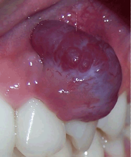
| A 14-year-old male patient was referred to our department with a complaint of growth in the mouth involving upper left anterior region which bled frequently and interfered with eating. His medical history was non-contributory. The patient noticed the growth two months back which was slightly smaller than the size at the time of presentation. Intra oral examination revealed a solitary growing exophytic, pedunculated lesion measuring 3x2x1 cm in the left upper anterior region attached to the marginal gingival interproximally between the left permanent lateral incisor and the canine (Figure 1). |
| |
|
|
Figure 1: Lobular capillary hemangioma involving the gingival. |
|
| |
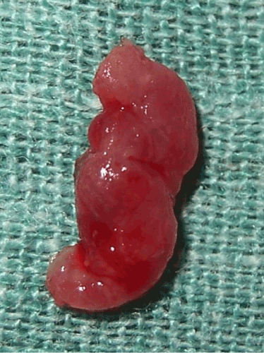
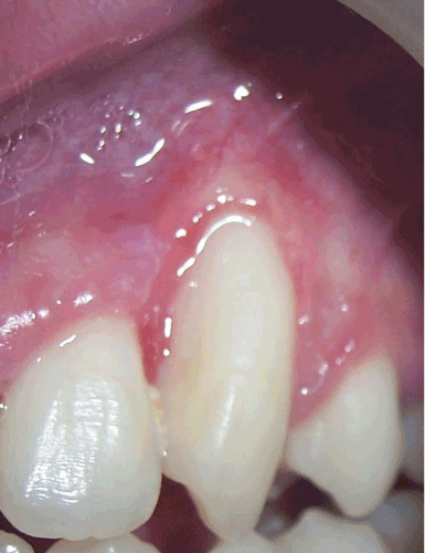
| The lesion was soft and edematous in consistency, tender with profuse bleeding on provocation. In addition, the patient had poor oral hygiene. After initial scaling and oral prophylactic treatment the lesion was excised and the gingival specimen was sent for histopathological examination (Figure 2). The healing was uneventful and patient was satisfied with the treatment outcome (Figure 3). |
| |
|
|
Figure 2: Specimen of the excised lesion. |
|
| |
|
|
Figure 3: Aesthetic treatment outcome after excising the lesion. |
|
| |
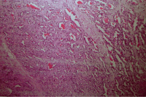
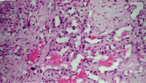
| The histopathologic examination revealed stratified squamous epithelium with acanthosis and irregular downward proliferation of rete pegs. There was a focal area of ulceration with underlying granulation tissue. The immediate sub-epithelium showed dense infiltration predominantly by plasma cells, neutrophils, lymphocytes and occasional eosinophils. There was presence of multinucleated giant cells of varying size in a stroma composed of ovoid and spindle shaped fibroblasts along with the presence of proliferating, some dilated and congested blood vessels (Figure 4,5). These findings were consistent with the histopathological diagnosis of pyogenic granuloma. |
| |
|
|
Figure 4: Histological slides showing the characteristic features of Lobular Capillary Hemangioma. |
|
| |
|
|
Figure 5: Histological slides showing the characteristic features of Lobular Capillary Hemangioma. |
|
| |
| Discussion |
| |
| Originally, pyogenic granulomas were believed to be botryomycotic infection which was transmitted from horse to man. Subsequently it was proposed that these lesions are caused due to some pyogenic bacteria like streptococci and staphylococci. However there is no evidence of any infectious organisms isolated from the lesions confirming the unlikely relation to any infection and hence the name is a misnomer. It is now largely agreed that pyogenic granuloma arises as a result of various stimuli such as low grade chronic irritation, trauma, hormonal imbalances or certain kinds of drugs. The tissues react in a characteristic manner resulting in overzealous proliferation of a vascular type of connective tissue [8]. The first case was reported in 1844 by Hullihen [9] and the term “pyogenic granuloma” or “granuloma pyogenicum” was coined only in 1904 by Hartzell [10]. |
| |
| In the oral cavity pyogenic granulomas show a striking predilection for the gingiva with interdental papillae being the most common site. They are more common in the maxillary anterior region than any other area in the mouth. Gingival irritation and inflammation that result from poor oral hygiene, dental plaque and calculus or over-hanging restorations may be the precipitating factors in many cases [7]. It is possible that micro-ulceration from these irritants in an already inflamed gingiva allows the ingress into the gingival connective tissue of low virulent oral micro-flora. Clinically the lesion is usually elevated, pedunculated or sessile, mass with smooth, sometimes lobulated and warty surface which can commonly show ulcerations covered with yellow fibrinous membrane. The colour ranges from deep red, reddish purple to pink depending on its duration and vascularity of the lesion. The lesion shows a tendency for hemorrhage either spontaneously or upon slight trauma. The lesion is painless and soft in consistency; although older lesions tend to become more collagenized and firm. The size of the lesion usually ranges between 0.5cm-2cm, and they may grow at an alarming rate reaching that size in just 4-7 days [5]. |
| |
| Although pyogenic granuloma can be diagnosed clinically with considerable accuracy, radiographic and histopathological investigations help in confirming the diagnosis. Radiographs are advised to rule out bony destructions suggestive of malignancy or to identify a foreign body [7]. Depending on its rate of proliferation and vascularity, there are 2 histological variants of pyogenic granuloma called Lobular Capillary Hemangioma (LCH type) and Nonlobular Capillary Hemangioma (non- LCH type) [8,11]. Differential diagnosis of PG includes parulis, peripheral giant cell granuloma, peripheral ossifying fibroma, hemangioma, peripheral fibroma, leiomyoma, hemangioendothelioma, hemangiopericytoma, bacillary angiomatosis, kaposis sarcoma, metastatic tumor, post extraction granuloma and pregnancy tumor. |
| |
| The treatment of pyogenic granuloma depends on the severity of the symptoms in the lesion. If it is small, painless and free of bleeding, clinical observation and follow up are advised [12]. If the lesions are huge, surgical excision and removal of causative irritants (plaque, calculus, foreign material, source of trauma) are among the choice of treatment. There is relatively high rate of recurrence (about 15%) after simple excision [7]. Other conventional surgical modalities for the treatment of pyogenic granuloma reported are cryosurgery in the form of either liquid nitrogen spray or a cryoprobe. Nd:YAG, CO2 and flash lamp pulsed dye lasers have also been used for the treatment of oral pyogenic granuloma [13]. |
| |
| Conclusion |
| |
| Lobular capillary hemangioma can present with unusual features. Although non neoplastic they can be present in uncommon site and with unusual size, therefore, proper management including diagnosis, treatment and further prevention is very important. Good oral hygiene, maintenance and regular follow- up can prevent recurrence of such lesions. |
| |
| |
| References |
| |
- Greenberg MS, Glick M (2003) Burkett’s Oral Medicine: Diagnosis and Treatment. (10stedn), BC Decker, Hamilton, Canada.
- Epivatianos A, Antoniades D, Zaraboukas T, Zairi E, Poulopoulos A, et al. (2005) Pyogenic granuloma of the oral cavity: comparative study of its clinicopathological and immunohistochemical features. Pathol Int 55: 391-397.
- Regezi JA, Sciubba JJ, Jordan RCK (2003) Oral Pathology: Clinical Pathological Considerations. (4stedn), WB Saunders company, Philadelphia, London, Toronto.
- Muench MG, Layton S, Wright JM (1992) Pyogenic granuloma associated with a natal tooth: case report. Pediatr Dent 14: 265-267.
- Ramirez K, Bruce G, Carpenter W (2002) Pyogenic granuloma: case report in a 9-year-old girl. Gen Dent 50: 280-281.
- Vilmann A, Vilmann P, Vilmann H (1986) Pyogenic granuloma: evaluation of oral conditions. Br J Oral Maxillofac Surg 24: 376-382.
- Amirchaghmaghi M, Falaki F, Mohtasham N, Mozafari PM (2008) Extragingival pyogenic granuloma: a case report. Cases J 1: 371.
- Shafer WG, Hyne MK, Lcvy HM (1983) Shafer's textbook of Oral Pathology. (4stedn), WB Saunders, Philadelphia, London, Toronto.
- Hullihen SP (1844) Case of aneurysm by anastomosis of the superior maxilla. Am J Dent Sc 4: 160-162.
- Hartzell ME (1904) Granuloma Pyogenicum. J. Cutan. Dis. Symph 22: 520-525.
- Neville BW, Damm DD, Allen CM, Bouquot JE (2002) Oral and Maxillofacial Pathology. (2stedn), WB Saunders, Philadelphia, London, Toronto.
- Sills ES, Zegarelli DJ, Hoschander MM, Strider WE (1996) Clinical diagnosis and management of hormonally responsive oral pregnancy tumor (pyogenic granuloma). J Reprod Med 41: 467-470.
- Jafarzadeh H, Sanatkhani M, Mohtasham N (2006) Oral pyogenic granuloma: a review. J Oral Sci 48: 167-175.
|
| |
| |