|
|
| Venkatachalapathy TS*, Sreeramulu PN and Nikhil S Shetty |
| Department of Surgery, Sri Devaraj Urs Medical College and Rl Jalappa Hospital and Research Centre, Tamaka, Kolar, Karnataka, India |
| *Corresponding authors: |
Venkatachalapathy TS
Department of Surgery
Sri Devaraj Urs Medical College and Rl Jalappa Hospital and Research Centre
Tamaka, Kolar, Karnataka
INDIA-563101
Tel: 8197507094
E-mail: drvenkey@hotmail.com |
|
| |
| Received March 28, 2012; Published August 22, 2012 |
| |
| Citation: Venkatachalapathy TS, Sreeramulu PN, Shetty NS (2012) Richter’s Hernia in Paraumbilical Region with Caecum and Ascending Colon as Content of Sac: a Case Report. 1: 266. doi:10.4172/scientificreports.266 |
| |
| Copyright: © 2012 Venkatachalapathy TS, et al. This is an open-access article distributed under the terms of the Creative Commons Attribution License, which permits unrestricted use, distribution, and reproduction in any medium, provided the original author and source are credited. |
| |
| Abstract |
| |
| A 12 year old boy presented with history of irreducible swelling in the Para umbilical region, no history of vomiting and abdominal distension. On examination non tender swelling in the Para umbilical region, cough impulse present and soft in consistency. The swelling is not reducible and there was no change in skin around the swelling. Patient underwent exploration of the hernia which revealed Richter’s hernia with normal caecum and ascending colon as content with bands around the mesentery. |
| |
| Introduction |
| |
| Richter’s hernia [1] may be defined as an abdominal hernia in which only part of the circumference of the bowel is entrapped and strangulated in the hernial orifice. The segment of the engaged bowel is nearly always the lower portion of the ileum, but any part of the intestinal tract, [2,3] from the stomach to the colon, including even the appendix, may become incarcerated [4]. |
| |
| The precondition for the formation of this particular hernia, as stated by Richter, is determined by the size and consistency of the hernial orifice: it must be big enough to ensnare the bowel wall, but small enough to prevent protrusion of an entire loop of the intestine, and the margin of the hernial ring must be firm or, in Richter’s words, “possess strong spring-force.” According to others, the presence of a tight constricting ring is a prerequisite for strangulation and compromised blood circulation, which finally leads to ischemia and gangrene of the involved bowel [5,6]. Richter’s hernias tend to progress more rapidly to gangrene than ordinary strangulated ones. This can be observed in the series of Horbach, who found 45 Richter’s hernias among 146 strangulated hernias [7,8]. In these 45, he found necrosis of the bowel wall in 31 (69%); among the 101 ordinary strangulated hernias, he found bowel necrosis in only 25 (25%). This may be explained not only by the firm constricting ring that exerts direct pressure on the bowel wall [9,10], but also by the anatomical peculiarity that, as a rule, it is the free border of the intestine opposite the mesentery with the predominance of terminal arterioles that is involved. It can also be explained by the time factor. In most cases, where less than two thirds of the circumference of the bowel wall is involved. The lumen of the gut remains free and an alarming intestinal obstruction is absent [11-13]. This insidious pathologic feature of Richter’s hernia often leads to late diagnosis or even misdiagnosis, thus allowing time for bowel necrosis to develop [14-16]. |
| |
|
|
| |
|
|
| |
|
|
| |
| Case Report |
| |
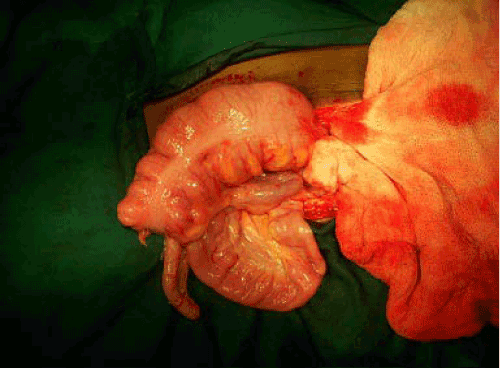
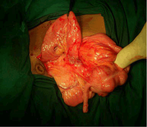
| We report a 12 year old boy presented with history of irreducible swelling in the Para umbilical region, no history of vomiting and abdominal distension. On examination non tender swelling in the Para umbilical region, cough impulse present and soft in consistency. The swelling is not reducible and there was no change in skin around the swelling. Blood investigations and other systemic examination was normal.USG (ultrasonography) revealed a Para umbilical hernia with enterocoele as the content noticed. On exploration there was cecum and ascending colon and appendix as the content of the sac was noticed. Ladd’s bands were seen at mesenteric region. Bowel viable hence contents reduced and defect closed with mayo’s repair. |
| |
| |
| References |
| |
- Tito WA, Allen WC (1989) Richter and Littre Hernia. In: Nyhus JB, Condon RE, eds. Hernia, (3rd edn) Philadelphia: Lippincott 305–310.
- Giokas G, Karakousis CP (1998) Richter hernia of the stomach. J Surg Oncol 69: 51-53.
- Duari M (1966) Strangulated femoral hernia--a Richter's type containing caecum and base of appendix. Postgrad Med J 42: 726-728.
- Rowe PH, Hunter-Craig C (1984) Richter's hernia in a direct inguinal sac in a female. J R Coll Surg Edinb 29: 264.
- Newerla GJ, Connally EF (1943) Gangrenous appendicitis in femoral hernia of Richter’s type. Am J Surg 61: 154–156.
- Pualwan F (1958) Operative aspects of Richter’s hernia. Surg Gynecol Obstet 106: 358–362.
- Keynes WM (1956) Richter's hernia. Surg Gynecol Obstet 103: 496-500.
- Treves F (1887) Richter’s Hernia or Partial Enterocele. Med Chir Trns 70: 149–167.
- Kadirov S, Sayfan J, Friedman S, Orda R (1996) Richter's hernia--a surgical pitfall. J Am Coll Surg 182: 60-62.
- Fückiger R, Huber A (1993) Die Richter’scheHernie. Der Chirurg 64: 822–826.
- Gillespie RW, Glas WW, Mertz GH, Musselman MM (1956) Richter's hernia; its etiology, recognition, and management. AMA Arch Surg 73: 590-594.
- Lyall D, Luomanen R (1948) Richter's hernia. Am J Surg 75: 828-833.
- Folimer HC (1966) Umbilical Richter’s hernia. Rocky Mt Med J 63: 61–62.
- Cook MD, Yarington CT, Jr. (1962) Strangulated Richter's hernia of the obturator canal: report of two cases. Am Pract Dig Treat 13: 115-118
- Naylor J (1978) Combination of Spigelian and Richter’s hernia: a case report. Am Surg 44: 750–752.
- Botsford TW (1977) Richter hernia in a sacral foramen: new site for richter hernia. Arch Surg 112: 304-305
- White CS (1912) Richter’s hernia. Surg Gynecol Obstet 14: 46–48.
|
| |
| |