|
|
| Montazeri M1, Rashidi N2*, Montazeri M3, Darabi J4 and Moghaddam KG5 |
| 1Cardiology Department, Tehran Heart Center Hospital, Tehran University of Medical Sciences; Tehran, Iran |
| 2Pulmonology Depatment, Shariati Hospital, Tehran University of Medical Sciences; Tehran, Iran |
| 3Pulmonology Department, Babol University of Medical Sciences; Babol, Iran |
| 4Radiology Department, Tehran university of Medical Sciences |
| 5Pulmonology Department, Shariati Hospital, Tehran University of Medical Sciences; Tehran, Iran |
| *Corresponding authors: |
Negin Rashidi
Pulmonology Department of Shariati Hospital
Tehran University of Medical Sciences (TUMS)
Northern Kargar Avenue
Tehran 1993643811, Iran
Fax: 0098-21-22976951
E-mail: rashidin@razi.tums.ac.ir |
|
| |
| Received August 28, 2012; Published August 23, 2012 |
| |
| Citation: Montazeri M, Rashidi N, Montazeri M, Darabi J, Moghaddam KG (2012) A Pulmonary Sequestration with Massive Hemoptysis. 1: 267. doi:10.4172/scientificreports.267 |
| |
| Copyright: © 2012 Montazeri M, et al. This is an open-access article distributed under the terms of the Creative Commons Attribution License, which permits unrestricted use, distribution, and reproduction in any medium, provided the original author and source are credited. |
| |
| Abstract |
| |
| Pulmonary sequestration is a rare malformation of the pulmonary parenchyma. Although pulmonary sequestration is rare finding but should be considered in patients with repeated episodes of bleeding. We describe a case of intralobar pulmonary sequestration found in a 27-year-old man admitted with massive hemoptysis and cough. He underwent lobectomy and achieved an excellent outcome. According to the potentially severe complications that they may cause, pulmonary sequestration should be removed whenever it is diagnosed; besides that repeated episodes of hemoptysis should be evaluated by more precise techniques such as ct angiography rather than broncoscopy. |
| |
| Keywords |
| |
| Intralobar Pulmonary Sequestration; Hemoptysis; Lobectomy |
| |
| Introduction |
| |
| Pulmonary sequestration (also known as bronchopulmonary sequestration) is a rare malformation of the pulmonary parenchyma that accounts for 0.15-6.4% of all congenital lung anomalies [1]. In pulmonary sequestration, a segment of lung parenchyma is separated from tracheobronchial tree, and is supplied by a systemic artery. Pulmonary sequestration usually appears enclosed in its own pleura (extralobar) or lies within the normal pulmonary visceral pleura (intralobar) [2]. Extralobar type presents early and is often diagnosed during infancy. Intralobar type either presents in early adulthood with repeated pulmonary infection or it might be an incidental findings [2,3]. The sequestration should be removed whenever it is diagnosed. Surgical resection is the only curative treatment for intrapulmonary sequestration. The most common surgeries are lobectomy, segmentectomy and simple mass excision [4]. We describe a case of intralobar pulmonary sequestration found in a 27-year-old man admitted with massive hemoptysis and cough. |
| |
| Case Report |
| |
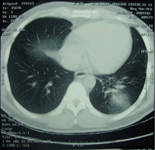
| A 27-year-old man admitted to our hospital with massive hemoptysis and cough. He did not complain of dyspnea or chest discomfort. At presentation he was afebrile, with BP=120/75 and pulse rate of 104 beat per minute. The patient was hospitalized three years ago with massive hemoptysis and cough. At that time, bronchoscopy was normal and the patient was discharged after improvement of symptoms. Bronchoscopy was performed for the second time and bleeding in the lower lobe of the left lung was seen. CT-Scan revealed a 63×41mm×42 mm dense consolidation in the peripheral area of lower lobe of left lung with air bronchogram (Figure 1). To determine the source of bleeding CT angiography was performed electively. CT Angiography showed a large artery arising from the descending aorta and supplying the left basal segment, and venous blood returning into the left inferior pulmonary vein (Figure 2). The Surgical procedure that was performed was left lower lobectomy. Postoperative histopathologic examinations confirmed that the lesion was an intralobar pulmonary sequestration. There was no malignant finding in the resected sample. The patient had an uneventful recovery and no complication was reported postoperatively. |
| |
|
|
Figure 1: A consolidation in the left lower lobe of left lung (hemorrhage in sequestrated lobe). |
|
| |
|
|
Figure 2: A large artery arising from descending aorta. |
|
| |
| Conclusion |
| |
| Pulmonary sequestration is a rare congenital malformation that may be presented with several features. Massive hemoptysis is one of these symptoms. Our patient chief complaint was several episodes of massive hemoptysis. In case report of Tamaki et al. [5], Wessels and Svolgaard [6] and Ichihashit et al. [7] hemoptysis was the main clinical feature of patients with pulmonary sequestration. In Zhang et al. survey [8] in 33 years evaluation, 42 cases of pulmonary sequestration were studied and in 10 (23.8%) patients, hemoptysis was the chief complaint. Arrabal-Sanchez et al. [9] reported two case of pulmonary sequestration that one of them (intralobar) was associated with hemoptysis. Other clinical features of pulmonary sequestration were recurrent pneumonia; shortness of breath, chest discomfort and cough [3] that cough was reported in our patient too. |
| |
| In our patient, bronchoscopy showed bleeding in the lower lobe of left lung but three years before recent hospitalization, with the same symptoms, bronchoscopy was normal. In Ichihashit et al. [7] the bronchoscopy in a patient with pulmonary sequestration was normal. According to these case reports, bronchoscopy results may be normal in patients with pulmonary sequestration. Therefore repeated episodes of hemoptysis should be evaluated by more precise techniques such as ct angiography rather than bronchoscopy. |
| |
| In our patient, ct angiography showed a large artery arising from the descending aorta and supplying the left basal segment that was similar to cases in Tamaki [5] and Ichihashit [7] case reports. Finding an artery connecting the cystic mass directly to the aorta distinguishes a pulmonary sequestration from a Congenital Cystic Adenomatoid Malformation (CCAM) [3]. |
| |
| In our patient the lesion was an intralobar pulmonary sequestration. Gezer et al. [3] reviewed 27 patients with pulmonary sequestration that 19 (70%) were intralobar pulmonary sequestration. In Gao et al. [4] 19 from 33 patients had intralobar pulmonary sequestration. |
| |
| The sequestration should be removed whenever it is diagnosed. The best treatment for pulmonary sequestration especially for intralobar type is lobectomy, as was performed in our patient. Segmentectomy and Simple Mass Excision are the others treatments that may be performed [4]. |
| |
| According to the potentially severe complications that they can cause, pulmonary sequestration should be removed whenever it is diagnosed. |
| |
| Conflict of Interest: None. |
| |
| |
| References |
| |
- Loscertales J, Congregado M, Arroyo A, Jimenez-Merchan R, Giron JC, et al. (2003) Treatment of pulmonary sequestration by Video-Assisted Thoracic Surgery (VATS). Surg Endosc 17: 1323.
- Au VW, Chan JK, Chan FL (1999) Pulmonary sequestration diagnosed by contrast enhanced three-dimensional MR angiography. Br J Radiol 72: 709-711.
- Gezer S, Tastepe I, Sirmali M, Findik G, Turut H, et al. (2007) Pulmonary sequestration: a single-institutional series composed of 27 cases. J Thorac Cardiovasc Surg 133: 955-959.
- Gao SG, Cheng GY, Sun KL, He J (2007) [Diagnosis and surgical treatment of pulmonary sequestration]. Zhonghua Yi Xue Za Zhi 87: 1616-1617.
- Tamaki T, Kusuyama Y, Matsuoka S, Nishimura O (1996) [A case of Pryce's type I pulmonary sequestration with hemoptysis and bruit]. Kyobu Geka 49: 1040-1043.
- Wessels J, Svolgaard PB (1998) [Hemoptysis with intralobar pulmonary sequestration]. Ugeskr Laeger 160: 6982-6983.
- Ichihashi T, Yamamura K, Ishida F, Seki M, Watanabe Y, et al. (1995) [A case report of pulmonary sequestration (Pryce I) associated with incomplete lobulation of the left lung]. Kyobu Geka 48: 1057-1060.
- Zhang L, Ding J, Jiang G (1998) [Diagnosis and treatment of pulmonary sequestration]. Zhonghua Jie He He Hu Xi Za Zhi 21: 675-677.
- Arrabal Sanchez R, Benitez Domenech A, Pages Navarrete C, Fernandez de Rota Avecilla A, Fernandez Bermudez JL (1998) [Pulmonary sequestration: 2 cases (intralobar and extralobar) in surgically treated adults]. Arch Bronconeumol 34: 45-47.
|
| |
| |