| Research Article |
Open Access |
|
| Praveen kumar M*, Suseelamma D, Saritha S and Lingaswamy V |
| Department of Anatomy, Kamineni Institute of Medical Sciences, Narketpally, Nalgonda, Andhra Pradesh, India |
| *Corresponding author: |
Praveen kumar M
Department of Anatomy
Kamineni Institute of Medical Sciences
Narketpally, Nalgonda
Andhra Pradesh, India
Tel: 9959750207
E-mail: praveenkumarmennakanti@gmail.com |
|
| |
| Received January 09, 2012; Published September 30, 2012 |
| |
| Citation: Kumar MP, Suseelamma D, Saritha S, Lingaswamy V (2012) Multiple Renal Vascular Variations. 1:334. doi:10.4172/scientificreports.334 |
| |
| Copyright: © 2012 Kumar MP et al., This is an open-access article distributed under the terms of the Creative Commons Attribution License, which permits unrestricted use, distribution, and reproduction in any medium, provided the original author and source are credited. |
| |
| Abstract |
| |
| The development of the renal vessels account for the fact of the complicate development of the kidney. The present study was under taken in 20 cadavers and 10 abortuses. |
| |
| I. Two cadavers showed accessory renal arteries and |
| |
| II. One male cadaver showed the following multiple renal vascular variations. |
| |
| - Accessory renal artery on right side, |
| |
| - Abnormal origin of Right testicular artery |
| |
| - Retro-aortic left renal vein were found. |
| |
| The knowledge of variations of the renal vessels forms an essential guide line for urosurgeon during the kidney transplantation and segmental resurrection. |
| |
| The attempt is made to discuss the embryological and clinical significance of the above variations observed. |
| |
| Keywords |
| |
| Accessory renal artery; Segmental resection; Retro aortic left renal vein (RALRV) |
| |
| Introduction |
| |
| Renal arteries branch laterally from the aorta just below the origin of the superior mesenteric artery. Both cross the corresponding crus of the diaphragm at right-angles to the aorta. The testicular arteries are two long slender vessels usually arise anteriorly from the abdominal aorta at the level of the second lumbar vertebra, a little inferior to the origin of renal arteries. Each passes inferolaterally under the parietal peritoneum into the pelvic cavity. The right testicular artery lies anterior to the inferior vena cava. It may originate from the renal artery or as a branch from a suprarenal or lumbar artery [1]. The large renal vein anterior to the renal arteries and open into the inferior vena cava almost at right angles. The left is three times longer than right and cross anterior to the aorta [1]. |
| |
| However published studies and reports about accessory renal artery occur approximately 30% [2]. The origin of left testicular artery from accessory renal artery is also a rare variation. Retro-aortic left renal vein is a rare and important variation related to the development process. Occurrence all these variation in a single cadaver are very rare. |
| |
| In this we emphasize the importance of accessory renal artery on right side, right testicular artery arising from the same accessory renal artery. Retro-aortic renal vein is not only of academic interest but may also be of practical importance in radiological examination as well as in surgical investigation. |
| |
| Materials |
| |
| 20 cadavers and 10 abortuses were studied during 2009-2011 at Kamineni institute of medical sciences, Narketpally. |
| |
| Methods |
| |
| Dissection method |
| |
| Observations |
| |
| The following variations found in |
| |
| I. Two cadavers showed the accessory renal arteries. |
| |
| II. An elderly male cadaver showed the following variations. |
| |
| |
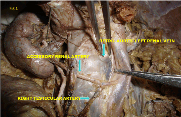
| - Accessory renal artery on right side arising from the abdominal aorta entering at the lower pole of the kidney in front of the ureter. |
| |
| - Right testicular artery originated from the same accessory renal artery (The origin of left testicular artery and the drainage of the right and left testicular veins are normal). |
| |
| - Presence left renal vein at back of the aorta (Retro-aortic left renal vein) (Figure 1). |
| |
|
|
Figure 1: Retro-aortic left renal vein |
|
| |
| Discussion |
| |
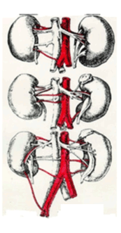
| In our case accessory renal artery is passing to the lower pole of the kidney in front of the ureter. Accessory renal arteries are not uncommon they are derived from the persistence of embryonic vessels that formed during the ascent of kidney. Kidneys develop in three stages of development pronephros, mesonephros and metanephros during this process the kidneys ascend from pelvic to the lumbar region. |
| |
| When the kidneys are situated in the pelvic cavity, they are supplied by the branches of common iliac arteries. While the kidneys ascend to lumbar region, their arterial supply also shifts from common iliac artery to abdominal aorta (Figure 2). Accessory renal arteries arise from the abdominal aorta either above or below the main renal artery and follow it to the hilum. It is important to be aware that accessory renal arteries are end arteries; therefore if an accessory is damaged, the part of kidney supplied by it is likely to become ischaemic [3]. |
| |
|
|
Figure 2: Common iliac artery to abdominal aorta |
|
| |
| Various chemical agents, growth/transcription factors and haemodynamic forces may all taken part in the selection and persistence of a particular congenital vascular channel. Embryonic signals that result in the formation of an accessory renal artery is yet unknown [4]. |
| |
| An accessory renal artery or leash arteries passing to the superior or inferior renal pole are possible. They are regarded as persistent embryonic lateral splanchnic arteries, accessory renal vessels to the inferior pole of the kidney cross anterior to the ureter and may, by its obstruction cause hydronephrosis. The urosurgeon should take care into account the origin of accessory renal artery when operating the lower pole of the kidney, segmental resection [5]. |
| |
| The rate of anatomical variation of testicular arteries has been reported to be 4.7% and their origin was either from unusually high level of aorta or from the renal artery [4]. |
| |
| Salve et al. has reported a variation in which the right testicular artery was arising from right aberrant artery (Accessory renal artery), in our case, right testicular artery arised from right accessory renal artery passing anterior to the inferior vena cava. |
| |
| These variations reported different regions of abdomen and pelvis are important not only in the view of development but also important for the surgeons dealing with kidney transplantation, obturator hernias and urogenital surgical procedures [6]. |
| |
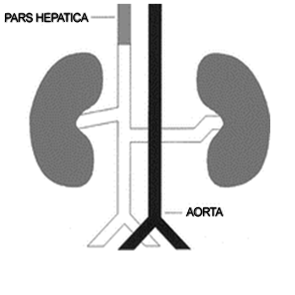
| As the anastomoses between the two supracardinal veins and the two subcardinal veins regress; a ventral renal vein is formed. During the normal formation of the left renal vein, the dorsal vessel degenerates and the ventral vessel become the renal veins. However, in the case of retro-aortic left renal vein, the ventral vessel degenerates and the dorsal vessel becomes the renal vein [7] (Figure 3). The published studies revealed the retro-aortic vein 0.8-7.1%. |
| |
|
|
| |
| Retro aortic left renal vein (RALRV) anomaly is a relatively uncommon condition which is present in our cadaver. Hemalatha [8] has reported the case study of a patient who had labile hypertension and left sided varicocoele, but no other clinical features of RALRV anomaly. |
| |
| Conclusion |
| |
| Out of 30 specimens |
| |
| I. Two cadavers showed accessory renal arteries and |
| |
| II. One male cadaver showed the fallowing multiple renal vascular variations. |
| |
| - Accessory renal artery on right side, |
| |
| - Abnormal origin of Right testicular artery from the above accessory renal Artery and |
| |
| - Retro-aortic left renal vein were found. |
| |
| |
| References |
| |
- Gray H, Bannister LH (1995) Grays anatomy. (38th edn), Churchill livingstone.
- Nathan H (1963) Aberrant renal artery producing developmental anomaly of kidney associated with unusual course of gonadal (ovarian) vessels. J Urol 89: 570-572.
- Nayak BS (2008) Multiple variations of the right renal vessels. Singapore Med J 49: e153-e155.
- Pai MM, Vadgaonkar R, Rai R, Nayak SR, Jiji PJ, et al. (2008) A cadaveric study of the testicular artery in the south indian population. Singapore Med J 49: 551-555.
- Kocabiyik N, Yalçin B, Kiliç C, Kirici Y, Ozan H (2005) Accessory renal arteries and an anomalous testicular artery of high origin. Gulhane Med J 47: 141-143.
- Bindhu S, Venunadhan A, Banu Z, Danesh S (2010) Multiple vascular variations in a single cadaver: A case report. Recent Research in Science and Technology 2:127-129.
- Sadler TW, Langman J (2010) Langmans’s medical embryology. (11th edn), Philadelphia: Lippincott William & Wilkins.
- Hemalatha K, Narayani R, Moorthy M, Korath MP, Jagadeesan K (2008) Retro-aortic left renal vein and hypertension. Bombay Hosp J 50: 1.
|
| |
| |