| Research Article |
Open Access |
|
| Oke AJ1*, Adeyemo AD2 and Oke AA3 |
| 1Department of Medical Microbiology, Bowen University Teaching Hospital, Ogbomoso, Nigeria |
| 2Department of Medical Microbiology, College of Medicine, University College Hospital, Ibadan, Nigeria |
| 3Department of Medical Microbiology, College of Health Sciences, Igbinedon University, Okada, Nigeria |
| *Corresponding authors: |
Dr. Oke AJ
Faculty of Basic Clinical Sciences
Bowen University Teaching Hospital
Ogbomoso, Nigeria
Tel: +2348034231902
E-mail: adefolaames@yahoo.com |
|
| |
| Received September 20, 2012; Published September 27, 2012 |
| |
| Citation: Oke AJ, Adeyemo AD, Oke AA (2012) Multidrug-Resistant Human Mycobacterium tuberculosis Strain Resisted Inactivation by 10% Formalin. 1:344. doi:10.4172/scientificreports.344 |
| |
| Copyright: © 2012 Oke AJ, et al. This is an open-access article distributed under the terms of the Creative Commons Attribution License, which permits unrestricted use, distribution, and reproduction in any medium, provided the original author and source are credited. |
| |
| Abstract |
| |
| Multidrug-resistant strain of Mycobacterium tuberculosis (MDR-Tb) and Multidrug -sensitive Mycobacterium tuberculosis (MDS-Tb) isolated from humans were injected subcutaneously into the guinea pig. One set of infected lungs of animals was fixed in 10% formalin pH 7.2, while the other set was treated with 75% ethanol, 2 hours prior to fixation in 10% formalin. |
| |
| After six months of fixation, MDS-Tb strains were completely inactivated by both methods of fixation. MDR-Tb strains resisted inactivation by 10% formalin, but were inactivated by the treatment with 75% ethanol 2 hours prior to 10% formalin fixation. |
| |
| Keywords |
| |
| Formalin; Ethanol; Inactivation; Mycobacterium tuberculosis |
| |
| Introduction |
| |
| Formalin is widely used to fix histological preparations, and as preservatives, in embalming solutions. It is an age-long practice in medical laboratories [1-3]. It is generally accepted that the risk of contracting tuberculosis is relatively high among Medical Laboratory workers and Pathologists. It is an assumption that once tissue is fixed in formalin, the risk for transmission and subsequent infection of Mycobacteria is greatly reduced, if not altogether eliminated [4-6]. Cadavers remain a practical teaching tool for Anatomists and Medical educators teaching gross anatomy. Infectious pathogens in cadavers that present particular risks include Mycobacterium tuberculosis, Hepatitis B and C, the AIDS virus HIV, and Prions that cause transmissible spongiform encephalopathies [7]. |
| |
| Recent studies have however, suggested that formalin does not effectively inactivate Mycobacterium tuberculosis in formalin-fixed tissue [4-13]. In this study we tried to test the inactivation ability of (a) 10% formalin fixation and (b) treatment of tissue with 75% ethanol, 2 hours prior to 10% formalin fixation. Multiple Drug Resistant (MDR) and Multiple Drugs Susceptible (MDS) strains of Mycobacterium tuberculosis were employed. |
| |
| Methods |
| |
| Official consent was obtained from the Head of Department, Chest Clinic, Baptist Medical Centre Ogbomoso. All subjects included in this study were volunteers. |
| |
| Sputa were collected from subjects at the Chest clinic of Baptist Medical Centre Ogbomoso. Samples were stained by Ziehl-Neelsen’s (ZN) technique for Acid Fast Bacilli (AFB). Positive AFB samples were cultured on Lowenstein-Jensen (L-J) medium before the commencement of multiple drug therapy [14,15]. The different drugs used at the clinic include; rifampicillin, isoniazid (INH), pyrazinamide and ethambutol. |
| |
| Primary growth on L-J was kept. Another round of sputa was collected from the same sets of subjects after the second and fifth months of directly observed multidrug therapy (DOT). The samples were stained. The positive AFB smears at the second and fifth month of DOT were considered to be MDR-Tb [16,17]. The negative AFB smears at the second and fifth months of DOT were considered MDSTb, and the primary growth from such samples were used as MDS-Tb. |
| |
| MDR-Tb sputa samples were cultured on L-J, and incubated. One set of animals was injected subcutaneously with the primary MDSTb growth, while another set was injected with the MDR-Tb growth [14]. Control animals were injected with sterile normal physiological saline. Animals were kept, fed on pellets and sterile water. They were watched for symptoms of disease, after which they were scarified and lungs excised. |
| |
| Parts of the lungs were processed for culture on L-J media to confirm infection in the animals. Parts of infected lung tissue were fixed in 75% ethanol for 2 hours and then in 10% formalin. Other parts of the lungs were fixed in 10% formalin pH 7.2. The fixed tissues were kept for 6 months. Lung tissues were processed for culture on L-J media, and incubated aerobically at 36°C. |
| |
| Results |
| |
| Fifty AFB-positive subjects were encountered in this study. Four (8%) were carrying MDR-Tb. |
| |
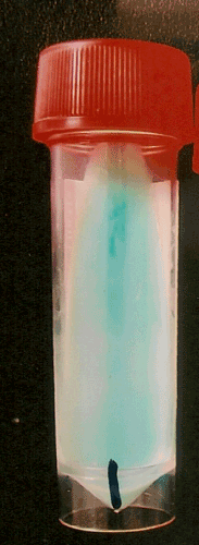
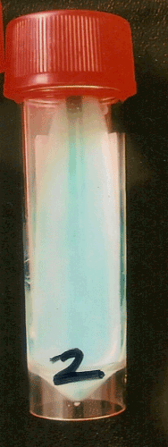
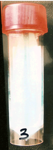
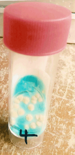
| The animals injected with mycobacteria cultures developed local swellings and weight-loss after one month, and mycobacteria were isolated from their lungs when cultured on L-J medium. There was no growth from the lung tissue of the control animals. There was no growth from the lungs of MDS-Tb infected animals in both fixation methods (Figures 1 and 2). In the MDR-Tb infected lungs, there was no growth in the 75% ethanol, 10% formalin fixation, but there was growth in the 10% formalin fixation (Figures 3 and 4). |
| |
|
|
Figure 1: MDS-Tb fixed in 10% formalin – No growth. |
|
| |
|
|
Figure 2: MDS-Tb fixed in 75% ethanol/10% formalin – No growth. |
|
| |
|
|
Figure 3: MDR-Tb fixed in 75% ethanol/10% formalin – No growth. |
|
| |
|
|
Figure 4: MDR-Tb fixed in 10% formalin-showing growth of Mycobacterium. |
|
| |
| This shows that both methods of fixation were able to inactivate MDS-Tb in lung tissue. MDR-Tb was not inactivated by 10% formalin fixation, but was inactivated by 75% ethanol/formalin fixation. |
| |
| |
| Discussion |
| |
| The results of our study show that treatment of lung tissue with alcohol before fixation in 10% formalin was able to inactivate Mycobacterium tuberculosis in tissue including MDR-strain that resisted 10% formalin. |
| |
| Environmental persistence of M. tuberculosis is subject of speculation. However, the reality that infected post mortem tissue can be a danger to Pathologists and embalmers has worrisome implications. A few experimental studies have demonstrated the ability of Mycobacterium tuberculosis to withstand exposure to embalming fluid and formalin. Gerston et al. succeeded in isolating live tubercle bacilli from 3 of 25 formalin fixed tissues. In a following report using a larger sample of 138 formalin-fixed lungs, they were able to obtain growth of Mycobacteria in three cases [5]. Treatment of tuberculosis is far from easy and frequently results in failure or even death [16-19]. The world is recording a high rate of death due to multiple-drug resistant tuberculosis. The results of workers suggested that there is a risk of contracting tuberculosis from tissue that has been fixed in formalin if aerosolization or accidental inoculation should occur [5]. In view of these, more effective fixative method should be employed to prevent occupational hazards to Medical Laboratory Scientists, Pathologists, Anatomists and Medical educators. |
| |
| It has been reported that alcohols, especially 70% ethanol, have rapid mycobactericidal activity [1], and a tuberculocidal fixation may be prepared with 10% formalin in 50% ethanol [10]. Treatment of tissues with 95% ethanol prior to fixation in 10% formalin has been suggested [20]. Multiple toxic effects of alcohols on structure and metabolism are suspected, primarily due to protein denaturation and coagulation. Alcohol had been reported to disrupt the amino acid-amino acid bond to form an amino acid-alcohol hydrogen bond [1]. |
| |
| The result of this investigations are in agreement with the suggestions of other workers for the use of alcohol in addition to the conventional 10% formalin fixation of tissues [10,20,21]. This is pertinent, more so now that more people die due to MDR-TB infections. |
| |
| Acknowledgement |
| |
| We acknowledge Mr. Festus Obaseki, TB Lab, UCH for his great assistance in culturing process; Head of Chest Clinic, Baptist Medical Centre Ogbomoso for giving consent; Mrs. Adigun C, for her assistance in sample collections; Mr. Tayo Adebayo who assisted in procuring the animals. |
| |
| |
| References |
| |
- McDonnell GE (2007) Antisepsis, disinfection, and sterilization: types, action and resistance. ASM Press Washington, DC, USA.
- Baker FJ, Silverton RE, Pallister CJ (1998) Introduction to Medical Laboratory Technology. (7thedn), Butterworth Heineman.
- Ochei J, Kolhalkar A (2007) Medical Laboratory science: Theory and Practice, Tata McGraw-Hill New Delhi, India.
- Gerston KF, Blumberg L, Gafoor H (1998) Viability of Mycobacteria in formalin-fixed tissue. Int J Tuberc Lung dis 2: 521.
- Gerston KF, Blumberg L, Tshabalala VA, Murray J (2004) Viability of mycobacteria in formalin-fixed lungs. Hum pathol 35: 571-575.
- Kappel TJ, Reinartz JJ, Schmid JL, Holter JJ, Azar MM (1996) The viability of Mycobacterium tuberculosis in formalin-fixed pulmonary autopsy tissue: review of the literature and brief report. Hum pathol 27: 1361-1364.
- Demiryürek D, Bayramoglu A, Ustaçelebi S (2002) Infective agents in fixed human cadavers: a brief review and suggested guidelines. Anat Rec 269: 194-197.
- Johnson BH, Davis F, Jamison RW, Marti WJ (1953) Viability and chemotherapeutic sensitivity of tubercle bacilli isolated from pulmonary and other lesions in embalmed bodies at autopsy; case report and clinico-pathologic correlations of cases studied to date. Dis Chest 23: 686-692.
- Meade GM, Steenken W Jr (1949) Viability of tubercle bacilli in embalmed human lung tissue. Am Rev Tuberc 59: 429-437.
- Vinnie DS, Mary R (2002) Does formaldehyde kill Myco tuberculosis? Tech Bull Histopath 2: 37-38.
- Lemma E, Zimhony O, Greenblatt CL, Koltunov V, Zylber MI, et al. (2008) Attempts to revive Mycobacterium tuberculosis from 300-year-old human mummies. FEMS Microbiol lett 283: 54-61.
- Sterling TR, Pope DS, Bishai WR, Harrington S, Gershon RR, et al. (2000) Transmission of Mycobacterium tuberculosis from a cadaver to an embalmer. N Engl J Med 342: 246-248.
- Lauzardo M, Lee P, Duncan H, Hale Y (2001) Transmission of Mycobacterium tuberculosis to a funeral director during routine embalming. Chest 119: 640-642
- Betty AF, Daniel FS, Alice SW (1998) Diagnostic Microbiology. (10thedn), Mosby.
- Gillies RR, Dodds TC (1973) Bacteriology Illustrated. (3rdedn), Churchill Livingstone.
- Olle-Goig JE (2006) Editorial: the treatment of multi-drug resistant tuberculosis--a return to the pre-antibiotic era? Trop Med Int Health 11: 1625–1628.
- García-García ML, Ponce de León A, Jiménez-Corona ME, Jiménez-Corona A, Palacios-Martínez M, et al. (2000) Clinical consequences and transmissibility of drug-resistant tuberculosis in Southern Mexico. Arch Intern Med 160: 630-636.
- Lockman S, Kruuner A, Binkin N, Levina K, Wang Y, et al. (2001) Clinical outcomes of Estonian patients with primary multidrug-resistant versus drug-susceptible tuberculosis. Clin Infect Dis 32: 373-380.
- Mukherjee JS, Rich ML, Socci AR, Joseph JK, Virú FA, et al. (2004) Programmes and principles in treatment of multidrug-resistant tuberculosis. Lancet 363: 474-481.
- Devlin R (2009) Tuberculosis autopsy, Clin Micronet.
- Benson AB (1990) Control of communicable diseases in Man. (15thedn), American Public Health Association, USA.
|
| |
| |