|
|
| Laith Mahmoud Abdulhadi* |
| Department of Prosthetic Dentistry, Faculty of Dentistry, University of Malaya, Kuala Lumpur, Malaysia |
| *Corresponding authors: |
Dr. Laith Mahmoud Abdulhadi
Department of Prosthetic Dentistry
Faculty of Dentistry University of Malaya
50603 Kuala Lumpur, Malaysia
Tel: +6-03-7967 7482
E-mail: laithabdulhadi@um.edu.my |
|
| |
| Received September 10, 2012; Published October 29, 2012 |
| |
| Citation: Abdulhadi LM (2012) Different Techniques for Palatal Augmentation in Partially Glossectomized Patients. A Report of Two Cases. 1:391. doi:10.4172/scientificreports.391 |
| |
| Copyright: © 2012 Abdulhadi LM. This is an open-access article distributed under the terms of the Creative Commons Attribution License, which permits unrestricted use, distribution, and reproduction in any medium, provided the original author and source are credited. |
| |
| Abstract |
| |
| Two patients with unilateral glossectomy due to removalof squamous cell carcinoma lumps on the lateral border were treated with partial and complete dentures palatal augmentation. Two different techniques were presented for constructing void palatal augmentation; one using functional impressiontechnique while, the other one was performed using functionally adapted modeling wax. The results were satisfactory for both techniques in the two patients regarding the enhancement of the tongue functions and communication. Therefore, the selection of which technique to be applied may depend only on the patient and practitioner preference. |
| |
| Keywords |
| |
| Palatal augmentation; Unilateral glossectomy prosthetic treatment |
| |
| Introduction |
| |
| During the past decades, an increased incidence of squamous cell carcinoma (SCC) of the tongue in young adults has been reported in several countries [1-4]. Some researches stated that male-to-female ratio was lower in young adults than in mature adults. Unlike the elder patients, many of young subjects had never used tobacco or over consumed alcohol. However, the time of exposure to these wellknown carcinogens in most cases was not long enough to contribute to malignant transformation. This suggests that other external or internal factors may play a role in the development of the disease in young adults. Hereditary may take part in this type of early-onset cancer, though reports in the literature are contradictory [5-7]. |
| |
| In healthy individuals, the tongue and the cheeks help to maintain the food on the occlusal table of the teeth. In addition, the tongue shapes the bolus of the food to prepare for deglutition. The tongue and the muscles of mouth floor then contract and elevate to squeeze the bolus against the palate to move it posteriorly. At the same time, the soft palate lifts to produce palatopharyngeal closure, and the muscles of the larynx contract to elevate the larynx and establish epiglottic closure of the larynx. In addition to its role in mastication and deglutition, the tongue is primary articulator for speech sounds. It functions in vowel productions by shaping the oral and pharyngeal cavities and in consonant production by restricting the oral cavity opening during articulation of linguo-velar, linguo-palatal, linguo-alveolar and linguo-dental consonant sounds [8]. Partial or total glossectomy results incompromised tongue’s functionsleading to a variable degree of deformity in speech articulation, deglutition and swallowing. Several types of prosthesis have been described to improve speech and swallowing in glossectomized patient. Their main function is to reshape the oral cavity, or to reduce its size, so that residual tongue can function more effectively in its oropharyngeal environment [9]. The purpose of this case report is to present two methods to augment the palate in two patients undergone partial hemiglossectomy. |
| |
| First case |
| |
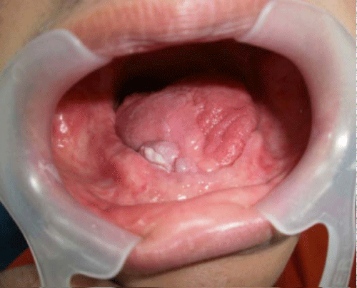
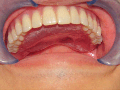
| A 31-year-old Chinese male was referred in June 2010 to the prosthetic dentistry department, for maxillary and mandibular complete dentures construction. The patient was operated onthe right sideborder of the tongue to eradicate a squamous cell carcinoma (SCC) lump. He underwent hemiglossectomy with right radical and left functional neck dissection and reconstruction with radial forearm flap in August 2008. Post surgically and prior to the radio and chemotherapy, his mouth was totally clearedof teeth due to caries. Medical history revealed that he was diabetic, hypertensivebut under medical control. He had never used prosthesis. Extra oral examination revealed asymmetrical face with postsurgical scarring on the neck area. The lips were competent with normal amplitude of mouth opening. The patient skeletal profile was class I. No temporomandibularjoint dysfunction signs and symptoms were detected except left joint click and mandible deviationtoward theright side during opening and closing movement. Intraorally, scar tissues was obliterating the right vestibule and extending to the remaining tissuesof the tongue causing limited movement. In addition, the maxillary and mandibular ridges were moderately resorbed (Figure 1). |
| |
|
|
Figure 1: The remaining of the tongue. |
|
| |
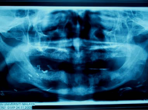
| Orthopantomogram (OPG) showed totally edentulous, moderately resorbedmaxillary and mandibular ridges (Figure 2). The treatment was to fabricate conventional maxillary and mandibular acrylic resin complete dentureswith unilateral palatal augmentation. Primary impression was made using stock tray with impression compound (Harvard, Berlin, Germany) for the maxillary arch. Irreversible hydrocolloid (Kromopan, Lascod, Illinois, and USA.) was used for the mandibular one. Final impression was made using close-fit custom tray constructed using light-cured acrylic resin materials (Plaque photo, W+P Dental, Hamburg, Germany) and zinc oxide/eugenol impression material (Impression paste, SS White, Gloucester, UK) formaxillary. While, silicone impression material (GC Exaflex regular, GC America, Alsip, USA)was used for the mandibular arch after border molding using tracing compound (Impression Compound Type I, Sds Kerr, USA). |
| |
|
|
Figure 2: The OPG of the patient after partial glossectomy. |
|
| |
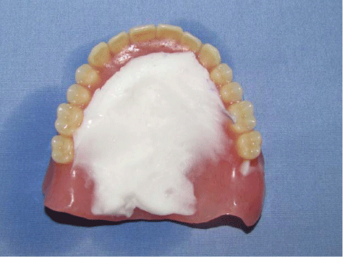
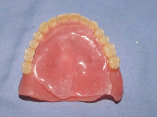
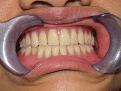
| Maxillomandibular relationship was recorded, teeth arranged, tried and then waxed dentures were processed using the ordinary wax elimination technique. The denture was issued and patient was instructed to use it for 1-2 weeks before palatal augmentation was done. The patient suffered difficult swallowing, saliva cooling, and speech problems. Therefore, thedecision was to augment the palate using functional impression technique with tissue conditioning material (Coe-Comfor, GC America Inc, Alsip, USA.) [10]. The material was added layer by layer to the palatal part of the denture and the patient was asked to swallow several times and pronounce some letters or phonemes (T, and D) [11]. Extra material was addeduntil optimum functions were achieved [12]. The patient was asked to use the denture for three days during loudly reading, swallowing, and eating soft diet in order to mold the tissue conditioning material to be compatible with the remaining tongue form and size during different functional movement. The denture was retrieved and a plaster matrix was fabricated to replace the tissue conditioning material with heat polymerized acrylic resin (Impact DEL-Dental Exports of London, UK) (Figure 3). On the plaster index, one layer of modeling wax was adapted to make a replica of the index. Then, the wax model was processed into heat cured acrylic resin using the normal wax elimination technique (Figure 4). The augmentation plate was polished, finished and then fixed in its place using autopolymerized acrylic resin (Satex, Metrodent, Paddock Huddersfield, UK). The denture rechecked inside the oral cavity for ease of swallowing, deglutition and speech functions and further enhancement may be made if required (Figures 5 and 6). |
| |
|
|
Figure 3: The molded palate. |
|
| |
|
|
Figure 4: The finished palate augmentation. |
|
| |
|
|
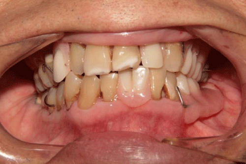
Figure 5: Maxillary denture with augmentation palate inside the mouth. |
|
| |
|
|
Figure 6: The maxillary and mandibular dentures inside the mouth. |
|
| |
| Second case |
| |
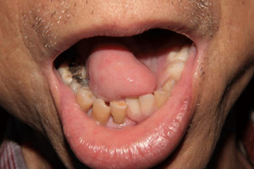
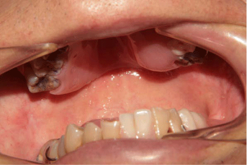
| A 72-year-old Chinese man was referred by the maxillofacial surgery department to find a solution for his continuous complaints regarding the inability to masticate the food, speech problems, xerostomia, and swallowing difficulty. He was operated to remove a lump on his left lateral tongue’s borderpartially after a diagnosis of (SCC) in 2006. The patient underwent right radical neck and left supraomohyoid neck dissections followed by radiotherapy (30 cycles). Extra oral examination revealed symmetrical face with postsurgical scarring on the neck. The lips were competent. The skeletal profile was class I. The maximum mouth opening was normal. Intra orally, scar tissues were obliterating the right vestibule and extending to join the remaining tissues of the tongue causing limited movement and resulted in a compromised speech, difficult mastication and deglutition (Figure 7). The maxillary arch was Class III Kennedy classification with two modifications, whereas the mandibular arch was Class II with one modification. The remaining teeth were badly carious due to multiple radiotherapy sessions. The decision was to construct acrylic resin maxillary removable partial denture (RPD) with palatal augmentationon the sideoftonguedefect to reduce the resulted space and to help reestablish easy physiologic contact between the mutilated tongue and the palate. The oral functions were enhanced dramatically as described by the patient. During the issuingtime, a palatal augmentation was done for the maxillary denture using alayer after layer of softened modeling wax until the patient experienced ease in swallowing using water for testing. The wax was softened and the patient asked to pronounce certain phonemes like B, K, T, S andsimilarly composingwords. A hallow augmentation palate was constructed to reduce the weight of the denture. Therefore, the same techniquefor making plaster index was used to fabricate the hollow palate either fromautopolymerizing acrylic resin (Satex, Metrodent, Paddock Huddersfield, UK) or preferably heat-cured acrylic resin. The augmenting acrylic piece was transferred into acrylic resin and fixed in its place on the palate using auto polymerizing acrylic resin (Figure 8). The patient explained his satisfactioneasy swallowing and better articulation of words after palatal remodeling (Figure 9). |
| |
|
|
Figure 7: The tongue defect of second patient. |
|
| |
|
|
Figure 8: The finished maxillary denture with augmentation. |
|
| |
|
|
Figure 9: The partial dentures inside the patient’s mouth. |
|
| |
| Discussion |
| |
| A minimum of 5 years of close monitoring is recommended for SCC patient due to high incidence of recurrence [11]. The Fabrication of prosthesis following hemiglossectomy is indicated when the patient experiences difficulty in speech and swallowing. The palatal augmentation prosthesis is a simple solution so that the tongue can reach again the palate during different functional movements. The prosthetic management of glossectomypatients improves mastication, swallowing, articulation of words and resonance. The food is easily redirected into the esophagus, and the remaining tissues are protected. Therefore, socialization is enhanced through improved communication in general [10]. In this report the two approachesproduced satisfied functional results for the two patients. In addition, the void type providedlighter prosthesis withoutauditable resonance. |
| |
| |
| References |
| |
- Myers JN, Elkins T, Roberts D, Byers RM (2000) Squamous cell carcinoma of the tongue in young adults: increasing incidence and factors that predict treatment outcomes. Otolaryngol Head Neck Surg 122: 44-51.
- Davis S, Severson RK (1987) Increasing incidence of cancer of the tongue in the United States among young adults. Lancet 2: 910-911.
- Schantz SP, Byers RM, Goepfert H (1988) Tobacco and cancer of the tongue in young adults. JAMA 259: 1943-1944.
- Shemen LJ, Klotz J, Schottenfeld D, Strong EW (1984) Increase of tongue cancer in young men. JAMA 252: 1857.
- Annertz K, Anderson H, Biörklund A, Möller T, Kantola S, et al. (2002) Incidence and survival of squamous cell carcinoma of the tongue in Scandinavia, with special reference to young adults. Int J Cancer 101: 95-99.
- Copper MP, Jovanovic A, Nauta JJ, Braakhuis BJ, de Vries N, et al. (1995) Role of genetic factors in the etiology of squamous cell carcinoma of the head and neck. Arch Otolaryngol Head Neck Surg 121: 157-160.
- Lichtenstein P, Holm N, Verkasalo P, Iliadou A, Kaprio J, et al. (2000) Environmental and heritable factors in the causation of cancer: analyses of cohorts of twins from Sweden, Denmark and Finland. N Engl J Med 343: 78–85.
- Mork J, Møller B, Glattre E (1999) Familial risk in head and neck squamous cell carcinoma diagnosed before the age of 45: a population-based study. Oral Oncol 35: 360-367.
- Aramany MA, Downs JA, Beery QC, Aslan Y (1982) Prosthodontic rehabilitation for glossectomy patients. J Prosthet Dent 48: 78-81.
- Hussein SZ (2000) Prosthodontic Rehabilitation Following Total and Partial Glossectomy, Clinical Maxillofacial Prosthetics edited by Thomas D Taylor. Quintessence Publising Co Inc, 205-213.
- Robbins KT, Bowman JB, Jacob RF (1987) Postglossectomy deglutitory and articulatory rehabilitation with palatal augmentation prostheses. Arch Otolaryngol Head Neck Surg 113: 1214-1218.
- Sessions DG, Spector GJ, Lenox J, Haughey B, Chao C, et al. (2002) Analysis of treatment results for oral tongue cancer. Laryngoscope 112: 616-25.
|
| |
| |