|
|
| Moutran R1* and Maatouk I2 |
| 1Dermatology department, Mount Lebanon hospital, Hazmieh, Lebanon |
| 2Dermatology department, Hotel Dieu de France hospital, Beirut, Lebanon |
| *Corresponding author: |
Maatouk I
Dermatology department
Mount Lebanon hospital
Hazmieh, Lebanon
Tel: +9613568968
Fax: +9611615300 ext. 9514
E-mail: ismaelmaatouk@yahoo.com |
|
| |
| Received September 05, 2012; Published October 29, 2012 |
| |
| Citation: Moutran R, Maatouk I (2012) Complication of a Red Tattoo. 1:410. doi:10.4172/scientificreports.410 |
| |
| Copyright: © 2012 Moutran R, et al. This is an open-access article distributed under the terms of the Creative Commons Attribution License, which permits unrestricted use, distribution, and reproduction in any medium, provided the original author and source are credited. |
| |
| Introduction |
| |
| Because of its wide popularity, tattooing is common in many different cultures and countries. Studies have reported various reactions to the salts and organic and inorganic compounds used in tattoos [1]. Among the most common reactions, those resulting from the red pigment deserve particular mention. We report a case of cutaneous reaction to the red pigment of a tattoo. |
| |
| Case |
| |
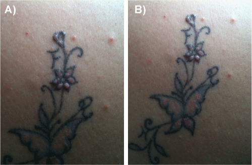
| A 52-year old female patient presented with non-pruriginous papulous lesions that had been present for 1 month on a tattoo performed 18 months previously on her right shoulder. The patient did not mention any skin nor internal disease and denied any medication/ drug consumption. She performed tattoos over her eyebrows 3 years ago without any complication. Examination revealed small papules located over the red pigment. The remaining blue part of the tattoo is normal (Figure 1a and 1b). Clinical exam was normal without any skin lesion elsewhere. Pulmonary and cardiac auscultation was normal. The patient refused to perform biopsy of the lesions. Her chest x-ray was normal. Blood test (blood formula, Angiotensin-converting enzyme, creatinin, urea, hepatic enzymes, electrolytes) was normal. Topical corticoid therapy was administered (Clobetasol propionate), resulting in a significant improvement in the appearance of the lesions. |
| |
|
|
Figure 1: Small papules located over the red pigment. The remaining blue part of the tattoo is normal. |
|
| |
| Discussion |
| |
| In Tahiti, the word tattow is translated as “painting in the skin” and the first published data on complications after tattooing return to Hutin in 1853 [2]. Currently, we know that the most common tattoo reactions are allergic contact dermatitis and lichenoid dermatitis principally due to the red pigment (mercury) [1-3]. The pigments in the tattoos contain many allergenic substances: hydrargyrum sulfide, cadmium sulfide, iron oxide, cobalt aluminate, manganese, chromium, and titanium. Allergic reactions to red, lilac, yellow, green, blue, and black tattoos have been described [2]. The tattoo pigments are often combined for obtaining different nuance. This makes extremely difficult the discovering of the cause for one allergic reaction. There are many cases concerning allergic reaction to the red dyes, which contain cinnabar (hydrargyrum sulfide). Allergic reactions include contact dermatitis, urticaria, photoinduced reactions (principally associated with yellow pigments) [2,4]. Other early complications may be due to infections caused by hepatitis (mainly HCV and HBV), HIV, herpes virus, poxvirus or papilloma virus, staphylococcus or streptococcus, mycobacterium, treponema, …[1,2]. Inflammatory reactions are less acute and include granulomatous reactions, pseudolymphoma and tumors. Lichenoid reactions occur in patients with previously known lichen planus. Koebner phenomenon may result in previously known lichen planus or psoriasis. |
| |
| Granulomatous reactions are seen weeks, months or years after tattoo. It is a foreign body reaction to the pigment and is associated with the use of mercury, chrome, cobalt and manganese [2]. It is generally limited to the colors of the tattoo and reflects a localized hypersensitivity reaction to the components of the pigments. Granulomatous reactions after tattooing are classified into 3 main categories-sarcoid granulomas, foreign body granulomas, and allergic granulomatous reactions [2]. Pure sarcoidal reactions are rare in tattoos. They generally occur as a reaction to the ochre dye, principally in silica-rich pigments. They may represent a nonspecific finding or a manifestation of systemic sarcoidosis [5-8]. There is no recommendation to perform any test to rule out a pulmonary sarcoidosis in this case [1]. |
| |
| The pseudolymphomatous reaction presents as hardened erythematous nodules or violaceous plaques that appear on the tattoo and at histology reveal germinative, infiltrated mixed-cell centers, principally in the superficial dermis and close to vessels. This type of reaction is more commonly associated with red, green and blue pigments [1,2]. Treatment with topical corticosteroid therapy is successful on granulomatous and lichenoid reactions. This is a case of cutaneous reaction to the red pigment of a tattoo. Since the patient refused the biopsy, we presume that it is a granulomatous reaction and treated with topical corticosteroid with partial regression of the papules. |
| |
| |
| |
| References |
| |
- Caucanas M, El Hayderi L, Lebas E, Richert B, Dezfoulian B, et al. (2011) Dermatological complications of temporary and indelible tattoos. Ann Dermatol Venereol 138: 161-162.
- Kazandjieva J, Tsankov N (2007) Tattoos: dermatological complications. Clin Dermatol 25: 375-382.
- Mortimer NJ, Chave TA, Johnston GA (2003) Red tattoo reactions. Clin Exp Dermatol 28: 508-510.
- Neri I, Giacomini F, Raone B, Patrizi A (2009) Generalized erythema multiforme after localized allergic dermatitis from dark henna tattoo. Pediatr Dermatol 26: 496.
- Ghorpade A (2006) Inoculation sarcoidal granulomas on blue-black tattoos in seven ladies. J Eur Acad Dermatol Venereol 20: 349-350.
- Antonovich DD, Callen JP (2005) Development of sarcoidosis in cosmetic tattoos. Arch Dermatol 141: 869-872.
- Landers MC, Skokan M, Law S, Storrs FJ (2005) Cutaneous and pulmonary sarcoidosis in association with tattoos. Cutis 75: 44-48.
- Collins P, Evans AT, Gray W, Levison DA (1994) Pulmonary sarcoidosis presenting as a granulomatous tattoo reaction. Br J Dermatol 130: 658-662.
|
| |
| |