| Research Article |
Open Access |
|
| Singh AK1, Ahmad Najam1, Pant AB2, Awasthi LP3*, Verma HN1 and Sharma NK3 |
| 1Virology Laboratory, Botany Department, Lucknow University, India |
| 2In vitro Toxicology Laboratory, Industrial Toxicology Research Centre, Lucknow, India |
| 3Department of Plant Pathology, Narendra Dev University of Agriculture and Technology, Faizabad, India |
| *Corresponding authors: |
L.P Awasthi
Professor & Head
Department of Plant Pathology
Narendra Dev University of Agriculture and Technology
Kumarganj, Faizabad-224 229, India
Tel: 91-5270-262012
Fax: 05270-262161
E-mail: lpawasthi06@yahoomail.com |
|
| Â |
| Received May 02, 2012; Published October 31, 2012 |
| Â |
| Citation: Singh AK, Najam A, Pant AB, Awasthi LP, Verma HN, et al. (2012) Modulatory Effect of Phytoproteins Isolated from Roots of Boerhaavia diffusa and Leaves of Clerodendrum aculeatum on Neoplastic Growth of Human Breast Cancer Cell Line. 1:429. doi:10.4172/scientificreports.429 |
| Â |
| Copyright: © 2012 Singh AK, et al. This is an open-access article distributed under the terms of the Creative Commons Attribution License, which permits unrestricted use, distribution, and reproduction in any medium, provided the original author and source are credited. |
| Â |
| Abstract |
| Â |
| The anticarcinogenic potential of phytoproteins isolated from B.diffusa and C. aculeatum were evaluated in MCF-7 cell lines obtained from human breast adenocarcinoma having the membrane receptor for estrogens and androgen. Cells were grown in Dulbecco's Modified Eagle's Medium (DMEM) supplemented with 10% fetal bovine serum, 0.1% antibiotic-antimycotic in a humidified atmosphere in 5% CO2 at 37°C. Monolayered batches of MCF- 7 cells showing cell viability more than 95% in Trypan Blue Dye exclusion test were used in all the experiments. Independent exposure of proteins of Boerhaavia diffusa showed a dose dependent reduction in cell growth at 0.1X, 1.0X and 10.0X concentrations in all the time intervals i.e., from 24 hr to 96 hr. However, 20.0X concentration were found to be cytotoxic. Isolated proteins of Clerodendrum aculeatum were found almost equally effective under experimental conditions. Interestingly, a synergistic effect on the growth inhibition (36.5 ± 3.1%) was observed on co-exposure of proteins of both the plants. However, mixture of proteins was also found to be cytotoxic at 20.0X concentration. The data of cell proliferation assay were also corresponding to the growth inhibition. Findings are suggestive that the combination of isolated proteins from Boerhaavia diffusa and Clerodendrum aculeatum have the potential of growth inhibition in MCF-7 cells under experimental conditions without having any cytotoxicity up to 10.0X concentrations and thus could be a useful anticancerous drug of future and will also be helpful for rural health care. |
| Â |
| Keywords |
| Â |
| Boerhaavia diffusa; Clerodendrum aculeatum; Anticarcinogenic proteins; MCF-7 cells; Human breast adenocarcinoma |
| Â |
| Introduction |
| Â |
| The roots of B. diffusa and leaves of C. aculeatum have shown to contain potent endogenous virus inhibitory proteins called as BD-SRIP and CA-SRIP, respectively [1-3]. These phytoproteins confer strong systemic resistance in several plants against a number of plant viruses [4-9]. Phytoproteins isolated from B. diffusa and C. aculeatum have molecular masses of 30 and 34 kDa, respectively. The phytoproteins are highly stable and could be purified [10,11]. |
| Â |
| Phytoproteins of B. diffusa and C. aculeatum have so far not been investigated for their antiviral activity against human and animal viral infections, though the studies on hepatoprotective activity of B. diffusa is documented [12]. Antiproliferative activity of alcoholic extract of B. diffusa in various cancerous cell lines has shown its potentiality to reduce the cell number without causing any cytotoxicity even at very high doses [13]. |
| Â |
| Trials similar to those performed for antiviral studies in plants were also conducted for anticancerous effects in animals [14]. The occurrence of phytoproteins possessing systemic resistance inducing property in plants makes the Boerhaavia diffusa plant very interesting compared to other virus inhibitor containing plants [10] which usually inhibit infections when mixed with virus inoculum in vitro. In view of the above facts the phytoproteins were tested against cancer cell line obtained from human breast cancer. |
| Â |
| The incidence of human breast cancer is continuously increasing. The situation is alarming particularly in developing and undeveloped nations where even basic necessities of livelihood are very scarce. Anticancer drugs are the only approach to minimize incidence of breast cancer and also reduce mortality associated with breast cancer. The drugs like tamoxifan effectively used in breast cancer therapy possess antiestrogenic activity. However, the production of these drugs is a costly affair and often unaffordable to people of poor nations. Therefore, identification and use of indigenous herbal medicines in cancer therapy can provide an alternative solution. Development of herbal medicine as an alternative cancer therapy has already attracted a great deal of attention not only due to their effectiveness but also due to their low toxicity and costs. |
| Â |
| Isolated and partially purified phytoproteins were screened for their anticancerous property using MCF7, a human breast adenocarcinoma cell line. The human breast cancer cell line such as MCF-7/adr (now known as NCI/ADR-RES) [15] T47D and ZR75 copiously express estrogen receptors (ERS). Estrogen after interaction with ERS initiates and regulates a cascade of events [16]. These events include expression of proteases and antiproteases, which prima facie regulate invasion and metastases. In this study we have used MCF-7/adr human breast cancer cell line. A MCF-7 cell line is a human breast adenocarcenoma which consists of epithelial cells isolated from pleural effusion of post menopausal female. |
| Â |
| Materials and Methods |
| Â |
| Sample collection |
| Â |
| The roots of B. diffusa were collected from fully mature plants growing in the district Kushinagar, Uttar Pradesh, India, whereas, the leaves of C. aculeatum were collected from plants growing as hedge in the Lucknow University campus, Lucknow. |
| Â |
| Isolation of proteins |
| Â |
| Extraction of proteins from roots of B. diffusa and leaves of C. aculeatum was carried out, essentially according to method described earlier [10,11] with desired modifications. The roots of B. diffusa were washed with distilled water, cut into small pieces, dried under natural sunlight and ground to fine powder in a grinder. The root powder was then mixed with 0.1 M phosphate buffer saline (PBS, pH 7.2) containing 0.1% 2-mercaptoethanol in the ratio of 1:5 and left overnight at 4°C. It was then filtered through two folds of muslin cloth and the filtrate so obtained was centrifuged at 8000 g for 15 minutes. In the supernatant ammonium sulphate 60% (w/v) was added with continuous stirring and left overnight at 4°C. The mixture was centrifuged at 8000 g for 15 minutes and the supernatant was discarded. The precipitate was retained and suspended in small amount of buffer [20 g fresh weight/ ml of 0.1M PBS (pH 7.2)] and then dialysed to obtain total protein fraction. The dialysed protein fraction was either diluted as per requirement or was concentrated through freeze-drying. Lyophilized protein was stored at -20°C. Essentially a similar protocol was adopted for obtaining a protein fraction from the leaves of C. aculeatum. |
| Â |
| Test material |
| Â |
| Lyophilised phytoproteins were used at different concentrations to evaluate their effectiveness against virus infestation in animals as well as the modulation in neoplastic growth of cancerous cells. Cells obtained from different cell lineage were used for different purpose. MCF-7/adr (now renamed as NCI/ADR-RE, human breast cancer) and L-929 (mouse fibroblast) cell lines were obtained from National Centre for Cell Science, Pune, India, and have been maintained in laboratory as per standard protocol. MCF-7 cells were cultured and maintained as per protocol [17]. Dulbecco’s Modified Eagel’s medium (DMEM) pH 7.4, supplemented with 10% fetal bovine serum and 10 mM EPES buffer and antibiotics (pencillin 100 units/ml, streptomycin 100 microgram/ml and gentamicin 60 μg/ml). L929, an adherent type mouse fibroblast cell line, recognized by ISO 10993 for the cytotoxicity assessment of biomedical devices and materials which are supposed to come in contact with human subject directly or through indirect means. L929 cells used in all the experiments of the present investigations were originally procured form NCCS, Pune and since then has been maintained at In Vitro Toxicology Laboratory, Industiral Toxicology Research Centre, Lucknow. |
| Â |
| Administration of phytoproteins |
| Â |
| The cytotoxic concentration of individual phytoproteins and their mixture was determined from a range of concentrations that had logarithmic relationship. Cells from L929 (mouse fibroblasts) lineage were used for this purpose. The phytoproteins used were directly added and thoroughly mixed with the medium recommended for the growth of these cells. Cytotoxicity was assessed using trypan blue exclusion test and MTT assay. Anticancerous activity was assessed in cells of MCF- 7/adr lineage. Only those doses that proved noncytotoxic were used. Individual phytoprotein or their mixtures were added directly to the medium. |
| Â |
| Cytotoxicity assay |
| Â |
| A range of concentrations of both the isolated proteins and their mixture were selected to ascertain the cytotoxic doses in L929, an adherent type of mouse fibroblast cell line recommended for cytotoxicity assessment by International and National Regulatory Agencies. Cytotoxicity was assessed using trypan blue dye exclusion test and MTT tetrazolium bromide salt assay. |
| Â |
| Trypan blue dye exclusion test |
| Â |
| The earlier method [18] with little modification was used to see the cell viability by assessing the loss of membrane integrity. In principle, the cells with damaged membrane allow the trypan blue dye to pass into the cytoplasm whereas undamaged cells are capable of dye exclusion. In brief, L929 cells (10,000) were seeded in 96-well tissue culture plates and incubated in the CO2 incubator for 24 hr, at 37°C for the proper attachment of the cells. Then the medium was precipitated and cells were exposed to different concentrations of test proteins (prepared in the complete MEM) for 24, 48, 72 and 96 hrs, under same conditions. The untreated set was also run simultaneously under identical conditions and served as control. By the end of exposure time, cells were trypsinized, centrifuged and pellet was re-suspended in the complete culture medium. Well-mixed cell suspension (0.8 ml) was added to a test tube already containing 0.2 ml of trypan blue (0.4%) and contents mixed by gentle shaking. In continuation, the dye-cell mixture was placed on the edge of both the chambers of the haemocytometer prefixed with the coverslip and allowed the cell suspension to fill the chambers by capillary action. The counting was made for unstained (viable cells) and stained cells (nonviable cells) in the four large corner squares in both counting chambers using field of 10X objective lens. The percent cell viability was calculated by deducting the number of stained cells from total number of cells counted (stained cells and unstained cells) over the haemocytometer. |
| Â |
| Mitochondrial activity by MTT assay |
| Â |
| The assay was done following the method of Pant et al. (2005) [19] with desired modifications. MTT assay provides an indication of mitochondrial integrity and activity, which is interpreted as a measure of cell viability. Viable cells absorb and reduce MTT a tetrazolium salt, 3-(4, 5-dimethylthiazol-2-yl)-2, 5 diphenyltetrazolium bromide in a mitochondrial-dependent reaction to yield a formazon product. The product accumulates within the cell since it cannot pass through the cell membrane. Upon addition of DMSO, isopropanol, or other suitable solvent, the product is solubilized and liberated and is readily quantified colorimetrically at 530 nm. Cells (10,000 in number) were seeded in 96-well tissue culture plates and incubated in the CO2 incubator for 24 hr at 37°C for the proper attachment of the cells. Then the medium was aspirated and cells were exposed to different concentrations of isolated proteins and their mixture for 24, 48, 72 and 96 hrs. MTT, 10 μl (5 mg/ml stock solution) in PBS, was added to every well and the culture plates incubated at 37°C. On completion of respective incubation periods, the reaction mixture was carefully taken out and 200 μl of DMSO was added to each well by pipetting up and down several times unless the content got homogenized. The cells grown in the medium only, without test compounds were treated as negative control and with 17 β-estradiol (1 μM, 0.1μM and 1nM) were treated as positive control. After 10 minutes, the color was read at 530 nm, using ELISA plate reader (BioRad, Tokyo, Japan). A standard curve was constructed at the start of every experiment. |
| Â |
| Cell proliferation assays |
| Â |
| Cell proliferation assay was used to assess the estrogenic and anti estrogenic properties of the phytoproteins. The assay detects both the magnitude of the agonist and antagonist effect on the cell number as well as the dose of agonist necessary to induce the proliferative effect of estradiol (agonist cancels the proliferative effect). Initially the cell number was quantified using electronic cell and particle counter. Cells (20,000 in number) were seeded in 24 well tissue culture plates and the cells were incubated in the CO2 incubator for 24 hrs, at 37°C for the adherence of the cells. Then the medium was aspirated and cells were exposed to different concentrations of the test substance. After 6 days the cells were trypsinized and counted in the coulter counter. The proliferation percentage was calculated with reference to positive and negative control. |
| Â |
| Results and Discussion |
| Â |
| Cytotoxicity studies |
| Â |
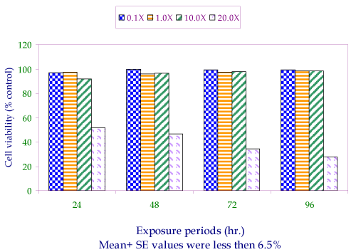
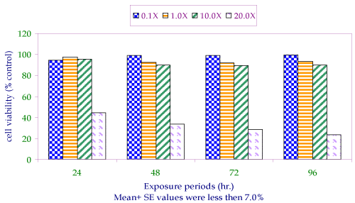
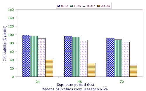
| All the doses used in the study were found to be non cytotoxic in L929 cells for an exposure upto 96 hr, except in case of 20X (10.0X+10.0X). Both the phytoproteins isolated from B. diffusa and C. aculeatum showed a time dependent decrease in cell viability, which lasted at 28.0 ± 2.16 and 23.4 ± 2.18 respectively by the end of incubation period i.e., 96 hr at 20X concentration. Cell viability was found more than 85% with respect to the untreated control for all the remaining doses used in the study and were considered as non cytotoxic doses (Figure 1-3). |
| Â |
|
|
Figure 1: Cell viability assay in L929, a mouse fibroblast (trypane blue dye exclusion test) post exposure to various concentrations of protein isolated from roots of Boerhaavia dffusa. |
|
| Â |
|
|
Figure 2: Cell viability assay in L929, a mouse fibroblast (trypane blue dye exclusion tset) post exposure to various concentrations of protein isolated from leaves of Clerodendrum aculeatum. |
|
| Â |
|
|
Figure 3: Cell viability in L929, a mouse fibroblast (trypane blue dye exclusion test) post exposure to various concentrations of mixture of proteins isolated from the roots of Boerhaavia diffusa and leaves of Clerodendrum aculealum. |
|
| Â |
| MTT assay |
| |
| Â |
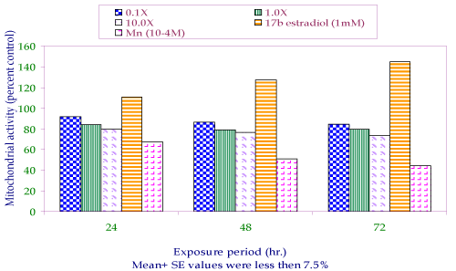
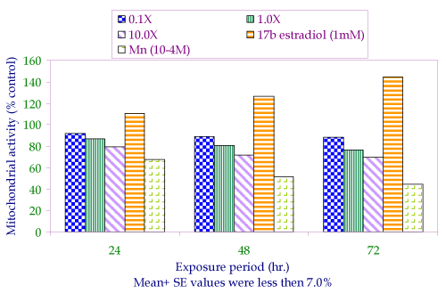
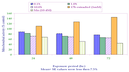
| MTT assay was done using MCF-7 cells to see the effect of test phytoproteins at various concentrations on the metabolic state of the cell and biomarker for cell growth commonly used to correspond the percent cell viability. The data of the MTT assay is well corresponding to the data of cytotoxicity in L929 and showing the same trend for all the concentrations used in the study. However, lower values of MTT assay in all the cases, might be due to the physiologically compromised state of few number of cells. In a dose and time dependent decreasing pattern of both the phytoproteins, the maximum reduction in the cell growth was 26.5 ± 2.18 and 30.1 ± 2.80 percent post 96 hr exposure for phytoproteins of B. diffusa and C. aculeatum respectively at 10X concentration. Interestingly, under the same concentration, the reduction was increased when the cells were exposed to the mixture of both the proteins i.e., 33.1 ± 3.07 percent at 96 hr (Figure 4-6). |
| Â |
|
|
Figure 4: Percent cell reduction in MCF-7 cells by MTT assay post exposure to varous concentrations of protein extracted from roots of Boerhaavia diffusa. |
|
| Â |
|
|
Figure 5: Percent cell reduction in MCF-7 cells by MTT assay post exposure to various concentration of protein extracted from leaves of Clerodendrum aculeatum. |
|
| Â |
|
|
Figure 6: Percent cell reduction in MCF-7 cells by MTT assay post exposure to various concentrations of proteins isolated from roots of Boerhaavia diffusa and leaves of Clerodendrum aculeatum. |
|
| Â |
| Proliferation Assay |
| Â |
| The purpose of this study was to ascertain the proliferative potential of cells in the absence and presence of E2 and to ascertain its regulatory effect of isolated phytoproteins from two medicinal plants (Table 1). The increase in cell numbers from 2.0×10⁴ (Day 0) at the time of plating to 10.21 ± 1.01 on Day 6 (100%) under the conditions of incubation confirms the normal proliferative potential of the cell line. Additionally, 1.0 μM, 0.1 μM and 1.0 nM concentrations of 17-β estradiol (E2) significantly upregulated the cell number by 123.5%, 67.0% and 48.2% respectively over Day 6 control. Like the results of MTT assay and dye exclusion test, the effect was dose dependent and a synergistic effect could be recorded when cells were exposed with the mixture of phytoproteins of both the plants i.e., 42.8% decrease in the cell number in comparison to control. 10X concentration of mixture proved maximum effective (42.8% decrease) followed by 10X of concentrations of individual phytoproteins (31.2% decrease in B. diffusa and 33.0 decrease in C. aculeatum) (Table 1). |
| Â |
|
|
Table 1: Antiproliferative effect of isolated phytoproteins in MCF-7 cells after six days of exposure. |
|
| Â |
| Morphological studies |
| Â |
| The cells were examined daily under a Leica Phase-contrast Inverted Microscope and photographed on Day 6, which marked the culmination of the experiment. The details of their morphological characteristics upon treatment with different concentrations and comparison with controls are as follows. (Figure 7a) depicts the control (without any treatment) cells on Day 6 with a typical oblong, polygonal appearance glued to the substratum in a flattened cobblestone fashion. Abundant cytoplasm was observed in the entire cell populace, which contained nuclei. Interspersed between the cells were occasional gap junctions signaling that the cells were in the replicative mode. The circumstantial presence of rounded dead cells was also noticed, floating in the medium in the background, which were removable upon replacement of the medium with fresh medium. Addition of 1.0 μM, 0.1 μM and 1.0 nM concentrations of 17-β estradiol (Figure 7b and 7c) displayed massive proliferative activity in a matter of 6 days confirming the steroid dependency of the cell line and resulted in a plethora of changes in the overall cellular architecture with more pronounced changes at the latter dosage. At 20X concentration of phytoprotein, the cells seemed reduced both in size and density as compared to the control (Figure 7d), whereas, with other concentrations used in the study, the cells were reduced in number but remain healthy with large amount of protoplasm in them (Figure 7e). Apparently, more reduction in the number of healthy cell was observed when cells were exposed to mixture of proteins of both the plants at 10X concentration. |
| Â |
| The idea to extend work on animal virus and cancer was based on those reports that indicated similarity in resistance responses among animals and plants. Some of the plant resistance genes (R genes) have been considered to be similar to Toll - like receptors that play important role in animal’s innate immunity. |
| Â |
| The various levels of the phytoproteins were either administered orally to the mice or were incorporated to the growth medium of different cell lines. In L 929 cell lines where cytotoxic level of the phytoproteins at logarithmically chosen levels was evaluated before carrying out antiviral tests on animal model, inhibitory effects or toxicity was observed at level of 20.0X. Neither of the phytoprotein at various chosen level of oral supply caused mortality nor induced any pathological symptoms in mice. Though the results obtained are contrasting particularly at higher concentrations - the differential activity may be related to the nature of the experimental material. In an intact animal, liver performs the function of detoxification of the various substances, so it is expected that the buildup of these phytoproteins in animals did not occur. However, in case of L929 fibroblast cell lines similar activity was lacking and therefore toxicity was noticed. |
| Â |
| The results in mice showing non toxic nature of phytoprotein were expected, as there is no report in literature on toxic effects associated with any of these plants, particularly in case of B. diffusa where root powder is used in the treatment of liver disorders [1]. In general, plant extracts show different degrees of toxicity. These toxic effects are most often dependent on the method of extraction. Higher toxicity has been reported in ethanol than in aqueous extracts [20-22]. Even in case of B. diffusa, the alcoholic extract of whole plant has not shown any toxicity effects even when administrated to an oral dose of 2 g/kg in mice [23]. For this reason and because the root powder of B. diffusa plant is taken by people either as decoction or infusion, water was preferred to prepare the extract. |
| Â |
| The roots of B. diffusa have therapeutic effects and are recommended for the treatment of anasarca, ascites and jaundice [24]. They have also reported to have diuretic, anti inflammatory, fibrinolytic nephrotic syndrome and anti-convulsant activities. These workers reported that administration of B. diffusa root extract was found to decrease activity of serum glutamate oxaloacetate transaminase (SGOT) and serum glutamate pyruvate transaminase. Some common anti-inflammatory medicinal plants such as Mentha spicata, Tephrosia purpurea, Cherranthus cheiri, Havsonia alba, Myrica nagri and Salix caprea have been shown to act as potent antioxidants as well as antitumor agents [25,26]. |
| Â |
| Though literature pertaining to medicinal importance of C. aculeatum is very rare to obtain, the phytoprotein was also tested for antitumorous and antiviral activity against animal virus because it possesses antiviral activity against plant viruses and has an action mechanism which is very similar to the antiviral activity of phytoprotein of B. diffusa root [2,6,9-11]. |
| Â |
| In antiproliferative studies conducted in MCF-7 human breast cancer cell line, both the phytoproteins reduced the cell proliferation rate. The antiproliferative activity was dose dependent. The phytoproteins of C. aculeatum, particularly at lower doses were more effective than phytoprotein of B. diffusa. When phytoproteins were used in combination, the effects were synergistic. It appears that the effector molecules act at different loci but these loci are constituent of same cell cycle control pathway. So when used in combination the action mechanism is coordinated to produce better effects than when used alone. The site of action C. aculeatum appears to be situated upstream to the site of action of B. diffusa. |
| Â |
| Recently, Mehrotra et al. (2002) [13] have shown that alcoholic extract of B. diffusa roots inhibited growth of several cancer cell lines of mouse and human origin. They have suggested that antiproliferative (anticancer) potential of extract may be due to the presence of ursolic acid. Ursolic acid is known to have anticancer activity and has also been isolated from B. diffusa [27]. It is suggested that the contrasting effects of phytoproteins of C. aculeatum on isolated cancer cell lines and on animals and ineffectiveness of phytoprotein of B. diffusa at lower doses may be either due to non appreciable absorption of phytoproteins in the alimentary canal or due to their degradation by liver enzymes; even a combination of these two factors could have occurred, causing a low concentration of phytoproteins in animal body than the effective dose. |
| Â |
| Conclusions |
| Â |
| Data of the present study provided strong evidence that the combinations of phytoproteins isolated from aqueous extract of B. diffusa and C. aculeatum have potential to regulate the growth of MCF-7 cells by inhibiting the cell proliferation under experimental conditions without having any cytotoxic effect upto a concentration of 10X and thus could be a anticancerous drug of future and may also be helpful for rural health care. However, to reach any firm conclusion and meaningful output, a series of in vitro, in vivo and clinical studies with the appropriate formulation and other characterization are required. |
| Â |
| Acknowledgements |
| Â |
| The authors are thankful Prof. Y. K. Gupta, Director, Industrial Toxicology Research Centre, Lucknow, India, for providing necessary laboratory faculties and keen interest in the study. |
| Â |
| |
| References |
| Â |
- Awasthi LP, Verma HN (2006) Boerhaavia diffusa -A wild herb with potent biological and antimicrobial properties. Asian Agri-History10: 55-68.
- Baranwal VK, Verma HN (1999) Antiviral phytoproteins as biocontrol agents for efficient management of plant virus diseases. In: Biocontrol Potential and Their Exploitation in Corp Pest and Disease Management (Edition Rajak RL and Rajeev K Upadhvay), 71-79.
- Verma HN, Srivastava S, Kumar D (1996) Induction of systemic resistance in plants against viruses by a basic protein from Clerodendrum aculeatumleaves. Phytopathology 86: 485-492.
- Awasthi LP (2009) Phytochemicals as inducers of systemic resistance against viral diseases in crops. Ind J Virol20: 24.
- Awasthi LP, Shyam Singh (2011) Management of ring spot disease of papaya through plant products. Indian Phytopath 62: 369-375.
- Singh S, Awasthi LP, Verma HN (2004) Prevention and control of yellow mosaic disease of mungbean through aqueous root extract of Boerhaavia diffusa. Ind Phytopathol 57: 303-307.
- Singh S, Awasthi LP (2004) Prevention of infection and spread of mungbean yellow mosaic virus (MYMV) on urdbean (Vigna mungo) through Boerhaavia diffusa root extract. Ind J Plant Pathol22: 50-55.
- Shyam Singh, Awasthi LP (2008) Protection of papaya (Carica papaya L.) against viral diseases through plant products. J Pl Dis Sci 3: 176-181.
- Yadav CP, Awasthi LP, Sanjay Singh (2009) Management of viral diseases of tomato through biopesticides: an ecofriendly approach.Ind J Virol20: 42.
- Verma HN, Awasthi LP (1980) Occurrence of a highly antiviral agent in plants treated withBoerhaavia diffusa inhibitor. Canadian Journal of Botany 58: 2141-2144.
- Verma HN, Baranwal VK, Srivastava S (1998) Antiviral substances of plant origin. In Plant virus disease control, edited by Hadidi A, Khetarpal RK, Kozenzava A, American Phytopathological Socity (APS) Press, St. Paul, Minnesota 154-162.
- Rawat AKS, Mehrotra S, Tripathi SC, Shome U (1997) Hepatoprotective activity of Boerhaavia diffusa L. roots-- a popular Indian ethnomedicine. JEthanopharmacol 56: 61-66.
- Mehrotra S, Singh VK, Agarwal SS, Maurya R, Srimal RC (2002) Antilymphoproliferative activity of ethanolic extract of Boerhaavia diffusa roots. Exp Mol Pathol72: 236-242.
- Singh AK, Najam A, Hussain MM, Verma HN, Awasthi LP (2009) Effect of phytoproteins isolated from roots of Boerhaavia diffusa and leaves of Clerodendrum aculeatum on the activity of Semliki forest Virus in albino mice model. Ind J Virol 20: 29-33.
- Liao CH, Pan SL, Guh JH, Chang YL, Pai HC, et al. (2005) Antitumor mechanism of evodiamine, a constituent from Chinese herb Evodiae fructs, in human multiple-drug resistant breast cancer NCI/ADR-RES cells in vitro and in vivo. Carcinogenesis 26: 968-975.
- Seo HS, Leclercq G (2002) Evaluation of potential implication of membrane estrogen binding sites on ERE-dependent transcriptional activity and intracellular estrogen receptor regulation in MCF-7 breast cancer cells. J Steroid Biochem Mol Biol 80: 109-123.
- Saxena AK, Ramchandani S, Dwivedi A, Sharma R, Bajpai VK, et al. (1995) Simplified cryopreservation of mammalian cell lines.In Vitro Cell Dev Biol Anim31: 326-329.
- Pant AB, Agarwal AK, Sharma VP, Seth PK (2001) In vitro cytotoxicity evaluation of plastic biomedicinal devices. Human Exp Toxicol20: 412-417.
- Pant VA, Sharma VP, Pant AB (2005) In vitro modulation in biological behaviour of cultured human periodontal related cells by platelet-rich plasma. J Ecophysiol Occupational Hlth5: 175-183.
- Abad MJ, Bermejo P, Gonzales E, Iglesias I, Irurzun A, et al. (1999) Antiviral activity of Bolivian plant extracts. Gen Pharmacol 32: 499-503.
- Bedoya LM, Sanchez-Palomino S, Abad MJ, Bermejo P, Alcami J (2001) Anti-HIV activity of medicinal plant extracts. J Ethnopharmacol77: 113-116.
- Goncalves JLS, Lopes RC, Oliveira DB, Costa SS, Miranda MM, et al. (2005) In vitro anti-rota virus activity of some medicinal plants used in Brazil against diarrhoea. JEthanopharmacol 99: 403-407.
- Chandan BK, Sharma AK, Anand KK (1991) Boerhaavia diffusa: a study of its hepatoprotective activity. JEthanopharmacol 31: 299-307.
- Chopra RN, Nayar SL, Chopra IC (1956)Glossary of lndian Medicinal Plants. C.S.I.R., New Delhi, India
- Alam A, Iqbal M, Saleem M, Ahmed S, Sultana S (2000) Myrica nagi attenuates the cumene hydroperoxide mediated cutaneous oxidative stress and toxicity in Swiss albino mice. Pharmacol Toxicol86: 209-214.
- Saleem M, Alam A, Sultana S (2001) Cheiranthus cheiri abrogates phorbol ester induce tumor promotion response in murine skin. Pharmaceutical Biology 39: 25-34.
- Sukhdev (2006) A selection of prime ayurvedic plant drugs. Ancient-modern concordance, Anamaya Publishers, New Delhi.
|
| Â |
| Â |