| Research Article |
Open Access |
|
| Neerja Puri*, Mahajan BB and Samarjit Kaur |
| Department of Dermatology and Pathology, GGS Medical College, Faridkot, 151203, India |
| *Corresponding author: |
Dr. Neerja Puri
# 626, phase II, Urban Estate
Dugri Road, Ludhiana, Punjab, India
Tel: 00919814616427
E-mail: neerjashu@rediffmail.com |
|
| Â |
| Received August 13, 2012; Published November 02, 2012 |
| Â |
| Citation: Puri N, Mahajan BB, Kaur S (2012) Clinicohistopathological Correlation of Psoriasis in Acute Exacerbation. 1:455. doi:10.4172/scientificreports.455 |
| Â |
| Copyright: © 2012 Puri N, et al. This is an open-access article distributed under the terms of the Creative Commons Attribution License, which permits unrestricted use, distribution, and reproduction in any medium, provided the original author and source are credited. |
| Â |
| Abstract |
| Â |
| Psoriasis is a genetically determined chronic disease of skin in which a number of biochemical and histopathological alterations take place during its course. An attempt was made to detect the histopathological changes during exacerbations and during the remission phase. Fifty patients of psoriasis were selected in acute exacerbation. Clinical examination, biochemical tests and skin biopsy was performed at the time of enrollment and at the end of eight weeks of treatment. With improvement of clinical picture; a shift of histopathological picture towards normal was recorded. |
| Â |
| Keywords |
| Â |
| Psoriasis; Histopathological; Infiltrate; Erythema; Munromicroabscess; Parakeratosis; Hyperkeratosis |
| Â |
| Introduction |
| Â |
| Psoriasis is a chronic recurrent papulosquamous disease characterized by epidermal hyperplasia [1]. Clinical diagnosis in a florid case shows well defined lesions with characteristic erythema and silvery white scales with auspitz sign [2]. But, there are occasions when while sitting in the outpatient department, one comes across a case with atypical or involuting or healed lesions with residual pigmented patches only and one is at ones wits ends about the diagnosis [3]. This fact prompted us to undertake the histopathological study of this disease in acute exacerbation and in the remission phase (resolving lesions). |
| Â |
| The cardinal histopathological features of psoriasis are a combination of the following: uniform parakeratosis, regular acanthosis in the mild intracellular and slight intercellular oedema, scattered mitosis of basal and prickle cells, papillomatosis, dilatation and tortuosity of the loops of capillaries and mild perivascular infiltration with lymphocytes [4]. Migration of leucocytes from capillaries in the tips of papillary bodies, through epidermis occurs to form the so called microabscesses, which is another confirmatory sign [5]. But all the characteristic features may not be present in one section alone. Also, it is important to note that dermis is affected earlier than epidermis in psoriasis. |
| Â |
| Materials and Methods |
| Â |
| We selected fifty patients from the outpatient department of dermatology, venereology and leprology. Detailed history, clinical examination, routine and biochemical investigations and histopathological examination was done in all the patients before the initiation of therapy and during the remission phase of the disease. |
| Â |
| The following patients were excluded from our study:- |
| Â |
| • Patients with impaired renal function or preexisting renal disease. |
| Â |
| • Patients with uncontrolled bacterial, viral or fungal infection. |
| Â |
| • Patients on concomitant use of hepatotoxic or nephrotoxic drugs for any other long standing cases. |
| Â |
| A written informed consent was taken from all the patients and prior approval from hospital ethics committee was taken. PASI score was recorded in all the patients before the initiation of the therapy and during the remission phase of the disease. |
| Â |
| Results |
| Â |
| The data was tabulated and the results of the study were analyzed statistically. Regarding the age distribution, it was seen that maximum number of patients (22%) were in age group of 51-60 years and the mean age of onset of psoriasis was 38.47 + 9.1 years. The male to female ratio was 1.94:1. Family history of psoriasis was positive in 8% patients. Regarding the incidence of different types of psoriasis, maximum percentage of patients was of psoriasis vulgaris (32%), followed by guttate posoriasis (28%). There were 16% patients of erythrodermic psoriasis, 12% patients of generalized pustular psoriasis and 12% patients were of palmoplantar psoriasis. Nail involvement was seen in 42% patients and psoriatic arthropathy was seen in 8% patients. |
| Â |
| Discussion |
| Â |
| Very few studies have been conducted regarding the histopathological changes in psoriasis before and after treatment. In our study, it was seen that in the active lesions, parakeratosis was more uniform, Munromicroabscesses were larger and more numerous [6]. There were dilated capillaries in the papillary bodies which were engorged with erythrocytes and leucocytes. The infiltrate was composed predominantly of polymorphonuclear leucocytes. In the psoriatic lesions in the resolving phase, there was no parakeratosis, no oedema or infiltration of cutis but, some degree of acanthosis and mild hyperkeratosis was evident [7]. Also, it was seen that the granular cell layer and parakeratosis seemed to alternate with each other i.e. whenever, there was frank, well developed parakeratosis, granular cell layer tended to be thinned or absent [8]. |
| Â |
| The incidence of munromicroabscess in psoriasis vulgaris group [9,10] was 43.75% and 57.14% in patients of guttate psoriasis [9,10]. The low incidence of microabscesses was explained on the basis of constant and pronounced exfoliation of the corneal layer [11]. Also, it was necessary to study multiple sections before microabscesses could be demonstrated [12]. Normally, all recognized histopathological features are not found in a well developed lesion of psoriasis. Therefore, the disparity between the present study and recognized histopathological features in a well developed lesion of psoriasis (i.e. absence of uniform parakeratosis, presence of normal stratum granulosum and normal thickness of suprapapillary plate in some specimens and lower incidence of munromicroabscesses) can be explained on the basis of biopsies of patients from different lesions [13]. |
| Â |
| For instance, some of them might have been quiescent or the regions of the lesions from where the biopsies had been taken were quiescent. This factor got reflected in their histopathological picture and was responsible for this disparity [14]. |
| Â |
| From the above discussion, it is evident that whereas the trend in the involuting lesions was its gradual reversal to normal histology in the epidermis (i.e. disappearance of parakeratosis, redevelopment of stratum granulosum, dimunition of acanthosis and attainment of normal thickness of stratum malpighii in the suprapapillary regions) there was no appreciable change in the histology of dermis [15]. |
| Â |
| Mild infiltrate, dilated capillaries and mild acanthosis, continued to persist in significant number of specimens even in this healed stage. Thus persistence of acanthosis, dilated capillaries and mononuclear infiltrate in the dermis in specimens from lesions taken which had healed even a year earlier, left first hypo or hyperpigmentation, though non specific, can be considered significant and sufficiently suggestive of previous lesion of psoriasis, especially when history of characteristic clinical features and other criteria like symmetrical distribution on extensor surface, seasonal variations and remissions and relapses is also present [16]. |
| Â |
| In our study, it was seen, that the healed lesions whether hyper or hypopigmented showed acanthosis, alongwith capillary dilatation and cellular infiltrate in dermis in a significant proportions of the specimens studied. It was also seen that apparently noninvolved skin of psoriatic, showed changes similar to those in the healed lesions of psoriasis in a significant number of specimens. |
| |
| Â |
| The occurrence and distribution of parakeratosis in a psoriatic lesion is definitely related to the degree of inflammatory reaction in the top of dermal papillae. This relationship between parakeratosis and papillae is not characteristic for psoriasis, but can also be seen in other dermatoses (eg. parapsoriasis and some eczema). In psoriasis this phenomenon is present most frequently and in many cases is very pronounced. This is attributed to the presence of thin suprapapillary parts of the malpighian layer together with a severe inflammatory reaction in the top of the papillae [17]. |
| Â |
| The clinically completely cleared lesions (Figures 1-6) (Tables 1-5) frequently showed histological changes and such lesions may relapse more rapidly than the lesions lacking histological changes. It was also found that the relapse rate was more in lesions treated with corticosteroids. |
| Â |
|
|
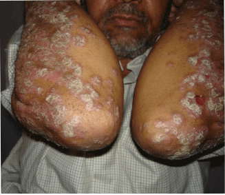
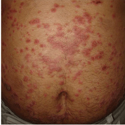
Figure 1a: Psoriasis vulgaris before treatment. |
|
| Â |
|
|
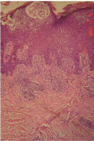
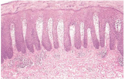
Figure 1b: Photomicrograph of psoriasis vulgaris before treatment showing Munromicroabcess, acanthosis, reterideges elongation and acute infiltration of dermis. |
|
| Â |
|
|
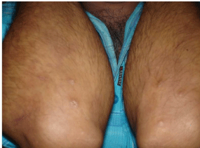
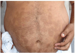
Figure 2a: Psoriasis vulgaris after treatment. |
|
| Â |
|
|
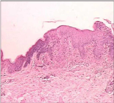
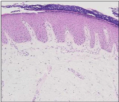
Figure 2b: Photomicrograph of psoriasis vulgaris after treatment. |
|
| Â |
|
|
Figure 3a: Guttate psoriasis in a 35 year old man before treatment. |
|
| Â |
|
|
Figure 3b: Photomicrograph of guttate psoriasis before treatment. |
|
| Â |
|
|
Figure 4a: Guttate psoriasis after treatment. |
|
| Â |
|
|
Figure 4b: Photomicrograph of guttate psoriasis after treatment. |
|
| Â |
|
|
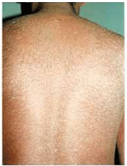
Figure 5a: Patient of psoriatic erythroderma before treatment. |
|
| Â |
|
|
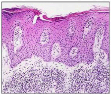
Figure 5b: Histopathology of psoriatic erythroderma before treatment. (H & E Stain). |
|
| Â |
|
|
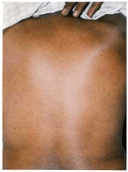
Figure 6: Patient of psoriatic erythroderma after treatment. |
|
| Â |
|
|
Table 1: Histopathological features of psoriasis vulgaris in acute phase and resolving phase. |
|
| Â |
|
|
Table 2: Histopathological features of Guttate psoriasis in acute & resolving phase. |
|
| Â |
|
|
Table 3: Histopathological features of psoriatic erythroderma in acute and resolving phase. |
|
| Â |
|
|
Table 4: Histopathological features of pustular psoriasis in acute and resolving phase. |
|
| Â |
|
|
Table5: Histopathological features of Palmoplantar psoriasis in acute and resolving phase. |
|
| Â |
| Conclusion |
| Â |
| The well developed lesions of psoriasis did not show all the characteristic histological features of psoriasis in all cases. The regression of histopathological changes of psoriatic lesions under treatment were first seen in the epidermis, especially in stratum corneum and granulosum and the changes in dermis persisted for a much longer time. The presence of histopathological changes in clinically cleared psoriatic lesions point to an important question, of how long a psoriatic skin lesion needs to be treated [18], in order to postpone recurrence as long as possible and achieve a permanent remission [19]. To conclude, the clinicopathological correlation is important especially to finally assess the outcome of the disease [20]. |
| Â |
| |
| References |
| Â |
- Guenther LC, Ortonne JP (2002) Pathophysiology of psoriasis: science behind therapy. J Cutan Med Surg 6: 2-7.
- Lebwohl M (2003) Psoriasis. Lancet 361: 1197-1204.
- Ashcroft DM, Wan Po AL, Williams HC, Griffiths CE (1999) Clinical measures of disease severity and outcome in psoriasis: a critical appraisal of their quality. Br J Dermatol 141: 185-191.
- Ragaz A, Ackerman AB (1979) Evolution, maturation, and regression of lesions of psoriasis. New observations and correlation of clinical and histologic findings. Am J Dermatopathol 1: 199-214.
- Bjerke JR, Krogh HK, Matre R (1978) Characterization of mononuclear cell infiltrates in psoriatic lesions. J Invest Dermatol 71: 340-343.
- Suurmond D (1965) Histologic changes in treated and untreated psoriatic lesions. Dermatologica 131: 357-366.
- Rowlands CG, Danby FW (2000) Histopathology of psoriasis treated with zinc pyrithione. Am J Dermatopathol 22: 272-276.
- Gordon M, Johnson WC (1967) Histopathology and histochemistry of psoriasis. I. The active lesion and clinically normal skin. Arch Dermatol 95: 402-407.
- Henseler T, Christophers E (1985) Psoriasis of early and late onset: characterization of two types of psoriasis vulgaris. J Am Acad Dermatol 13: 450-456.
- Brody I (1962) The ultrastructure of the epidermis in psoriasis vulgaris as revealed by electron microscopy: 1. The dermo-epidermal junction and the stratum basale in parakeratosis without keratohyalin. J Ultrastruc Res 6: 304-367.
- Munro JW, Montgomery H (1943) Histopathologic study of psoriasis. Arch Dermat and Syph 48:479.
- HELWIG EB (1958) Pathology of psoriasis. Ann N Y Acad Sci 73: 924-935.
- Madden JF (1941) Histologic studies of uninvolved skin of patients with psoriasis. Arch Dermatol 44 : 655-664.
- Artazaryan V, vetisyn A (1980) Immunomorphological and histopathological characteristics of skin in patients with psoriasis. Arch Pathol 42: 60-62.
- Zip C, Murray S, Walsh NM (1993) The specificity of histopathology in erythroderma. J Cutan Pathol 20: 393-398.
- Tomasini C, Aloi F, Solaroli C, Pippione M (1997) Psoriatic erythroderma: a histopathologic study of forty-five patients. Dermatology 194: 102-106.
- Boyd AS, Menter A (1989) Erythrodermic psoriasis. Precipitating factors, course, and prognosis in 50 patients. J Am Acad Dermatol 21: 985-991.
- Stern RS, Nijsten T, Feldman SR, Margolis DJ, Rolstad T (2004) Psoriasis is common, carries a substantial burden even when not extensive, and is associated with widespread treatment dissatisfaction. J Investig Dermatol Symp Proc 9: 136-139.
- Bowcock AM, Barker JN (2003) Genetics of psoriasis: the potential impact on new therapies. J Am Acad Dermatol 49: S51-56.
- Feldman SR, Fleischer AB Jr, Reboussin DM, Rapp SR, Exum ML, et al. (1996). The self-administered psoriasis area and severity index is valid and reliable. J Invest Dermatol 106: 183-186.
|
| Â |
| Â |